 Movie
Movie Controller

[English] 日本語
 Yorodumi
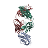
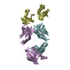
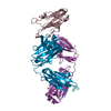
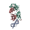
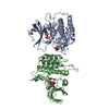
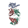
- PDB-1ira: COMPLEX OF THE INTERLEUKIN-1 RECEPTOR WITH THE INTERLEUKIN-1 RECE... -
+ Open data

- Basic information
| Entry | Database: PDB / ID: 1ira | ||||||
|---|---|---|---|---|---|---|---|
| Title | COMPLEX OF THE INTERLEUKIN-1 RECEPTOR WITH THE INTERLEUKIN-1 RECEPTOR ANTAGONIST (IL1RA) | ||||||
 Components Components |
| ||||||
 Keywords Keywords | COMPLEX (CYTOKINE RECEPTOR/ANTAGONIST) / CYTOKINE RECEPTOR / RECEPTOR ANTAGONIST / IMMUNOGLOBULIN FOLD / COMPLEX (CYTOKINE RECEPTOR-ANTAGONIST) / COMPLEX (CYTOKINE RECEPTOR-ANTAGONIST) complex | ||||||
| Function / homology |  Function and homology information Function and homology informationinterleukin-1 type I receptor antagonist activity / interleukin-1 type II receptor antagonist activity / positive regulation of interleukin-1-mediated signaling pathway / interleukin-1, type I receptor binding / interleukin-1 receptor antagonist activity / interleukin-1, type I, activating receptor activity / positive regulation of neutrophil extravasation / interleukin-1 receptor activity / interleukin-1, type II receptor binding / interleukin-1 binding ...interleukin-1 type I receptor antagonist activity / interleukin-1 type II receptor antagonist activity / positive regulation of interleukin-1-mediated signaling pathway / interleukin-1, type I receptor binding / interleukin-1 receptor antagonist activity / interleukin-1, type I, activating receptor activity / positive regulation of neutrophil extravasation / interleukin-1 receptor activity / interleukin-1, type II receptor binding / interleukin-1 binding / negative regulation of interleukin-1-mediated signaling pathway / smooth muscle adaptation / positive regulation of T-helper 1 cell cytokine production / negative regulation of heterotypic cell-cell adhesion / positive regulation of platelet-derived growth factor receptor signaling pathway / ADP-ribosyl cyclase/cyclic ADP-ribose hydrolase / NAD+ nucleosidase activity, cyclic ADP-ribose generating / platelet-derived growth factor receptor binding / interleukin-1 receptor binding / interleukin-1-mediated signaling pathway / insulin secretion / Interleukin-10 signaling / response to interleukin-1 / response to glucocorticoid / cytokine activity / acute-phase response / lipid metabolic process / positive regulation of type II interferon production / Interleukin-1 signaling / transmembrane signaling receptor activity / protease binding / regulation of inflammatory response / Potential therapeutics for SARS / positive regulation of canonical NF-kappaB signal transduction / cell surface receptor signaling pathway / positive regulation of phosphatidylinositol 3-kinase/protein kinase B signal transduction / immune response / inflammatory response / external side of plasma membrane / centrosome / cell surface / : / extracellular exosome / extracellular region / nucleoplasm / membrane / plasma membrane / cytosol Similarity search - Function | ||||||
| Biological species |  Homo sapiens (human) Homo sapiens (human) | ||||||
| Method |  X-RAY DIFFRACTION / MIR, MOLECULAR REPLACEMENT / Resolution: 2.7 Å X-RAY DIFFRACTION / MIR, MOLECULAR REPLACEMENT / Resolution: 2.7 Å | ||||||
 Authors Authors | Schreuder, H.A. / Tardif, C. / Tramp-Kalmeyer, S. / Soffientini, A. / Sarubbi, E. / Akeson, A. / Bowlin, T. / Yanofsky, S. / Barrett, R.W. | ||||||
 Citation Citation | Journal: Nature / Year: 1997 Title: A new cytokine-receptor binding mode revealed by the crystal structure of the IL-1 receptor with an antagonist. Authors: Schreuder, H. / Tardif, C. / Trump-Kallmeyer, S. / Soffientini, A. / Sarubbi, E. / Akeson, A. / Bowlin, T. / Yanofsky, S. / Barrett, R.W. #1: Journal: FEBS Lett. / Year: 1995Title: Crystals of Soluble Interleukin-1 Receptor Complexed with its Natural Antagonist Reveal a 1:1 Receptor-Ligand Complex Authors: Schreuder, H. / Tardif, C. / Soffientini, A. / Sarubbi, E. / Akeson, A. / Bowlin, T. / Yanofsky, S. / Barrett, R.W. #2: Journal: Eur.J.Biochem. / Year: 1995Title: Refined Crystal Structure of the Interleukin-1 Receptor Antagonist. Presence of a Disulfide Link and a Cis-Proline Authors: Schreuder, H.A. / Rondeau, J.M. / Tardif, C. / Soffientini, A. / Sarubbi, E. / Akeson, A. / Bowlin, T.L. / Yanofsky, S. / Barrett, R.W. | ||||||
| History |
|
- Structure visualization
| Structure viewer | Molecule: MolmilJmol/JSmol |
|---|
- Downloads & links
-Download
| PDBx/mmCIF format | 1ira.cif.gz | 108.7 KB | Display | PDBx/mmCIF format |
|---|---|---|---|---|
| PDB format | pdb1ira.ent.gz | 82.9 KB | Display | PDB format |
| PDBx/mmJSON format | 1ira.json.gz | Tree view | PDBx/mmJSON format | |
| Others |  Other downloads Other downloads |
-Validation report
| Arichive directory | https://data.pdbj.org/pub/pdb/validation_reports/ir/1iraftp://data.pdbj.org/pub/pdb/validation_reports/ir/1ira | HTTPS FTP |
|---|
-Related structure data
| Related structure data | 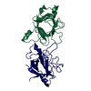 1ilrS  2cd4 S: Starting model for refinement |
|---|---|
| Similar structure data |


-Links
 PDBj
PDBj


- Assembly
| Deposited unit | 
| ||||||||
|---|---|---|---|---|---|---|---|---|---|
| 1 |
| ||||||||
| Unit cell |
|
-Components
| #1: Protein | Mass: 17145.406 Da / Num. of mol.: 1 Source method: isolated from a genetically manipulated source Source: (gene. exp.) Homo sapiens (human) / Cell line: SF9 / Plasmid: TAC-BSP / Production host:  | ||||
|---|---|---|---|---|---|
| #2: Protein | Mass: 36741.734 Da / Num. of mol.: 1 / Fragment: TYPE I RECEPTOR, EXTRACELLULAR DOMAINS Source method: isolated from a genetically manipulated source Source: (gene. exp.) Homo sapiens (human) / Cell line: SF9 / Plasmid: PVL1392 / Cell line (production host): SF9 / Production host:   Spodoptera frugiperda (fall armyworm) / References: UniProt: P14778 Spodoptera frugiperda (fall armyworm) / References: UniProt: P14778 | ||||
| #3: Sugar | ChemComp-NAG /   Type: D-saccharide, beta linking / Mass: 221.208 Da / Num. of mol.: 4 Type: D-saccharide, beta linking / Mass: 221.208 Da / Num. of mol.: 4Source method: isolated from a genetically manipulated source Formula: C8H15NO6 #4: Water | ChemComp-HOH / |  Mass: 18.015 Da / Num. of mol.: 86 / Source method: isolated from a natural source / Formula: H2O Mass: 18.015 Da / Num. of mol.: 86 / Source method: isolated from a natural source / Formula: H2OHas protein modification | Y | |
-Experimental details
-Experiment
| Experiment | Method: X-RAY DIFFRACTION / Number of used crystals: 1 |
|---|
- Sample preparation
| Crystal | Density Matthews: 2.7 Å3/Da / Density % sol: 54 % | |||||||||||||||
|---|---|---|---|---|---|---|---|---|---|---|---|---|---|---|---|---|
| Crystal grow | Method: vapor diffusion, hanging drop / pH: 7 Details: THE COMPLEX OF THE IL-1 RECEPTOR WITH THE ANTAGONIST WAS PREPARED BY MIXING A SOLUTION OF IL-1 RECEPTOR IN 50 MM TRIS (PH 7.5) AND 150 MM NACL WITH A SOLUTION OF IL1RA IN THE SAME BUFFER. ...Details: THE COMPLEX OF THE IL-1 RECEPTOR WITH THE ANTAGONIST WAS PREPARED BY MIXING A SOLUTION OF IL-1 RECEPTOR IN 50 MM TRIS (PH 7.5) AND 150 MM NACL WITH A SOLUTION OF IL1RA IN THE SAME BUFFER. THE COMPLEX WAS CRYSTALLIZED USING THE HANGING DROP METHOD. THE DROPS WERE PREPARED BY MIXING 4 UL OF THE PROTEIN SOLUTION WITH 1 UL OF RESERVOIR SOLUTION, CONTAINING 30% (W/V) PEG 3350, 400 MM MGCL2 IN 100 MM MOPS BUFFER (PH 7.0)., vapor diffusion - hanging drop | |||||||||||||||
| Crystal grow | *PLUS Temperature: 23 ℃ / pH: 8.8 / Method: vapor diffusion | |||||||||||||||
| Components of the solutions | *PLUS
|
-Data collection
| Diffraction | Mean temperature: 296 K |
|---|---|
| Diffraction source | Source: ROTATING ANODE / Type: MACSCIENCE M18X / Wavelength: 1.5418 |
| Detector | Type: SIEMENS / Detector: AREA DETECTOR / Date: Sep 1, 1994 / Details: COLLIMATOR |
| Radiation | Monochromator: GRAPHITE(002) / Monochromatic (M) / Laue (L): M / Scattering type: x-ray |
| Radiation wavelength | Wavelength: 1.5418 Å / Relative weight: 1 |
| Reflection | Resolution: 2.7→60 Å / Num. obs: 15631 / % possible obs: 97.2 % / Redundancy: 4.7 % / Rsym value: 0.06 |
| Reflection shell | Resolution: 2.7→2.8 Å / Redundancy: 3.1 % / Rsym value: 0.325 / % possible all: 85.3 |
| Reflection | *PLUS Num. measured all: 73407 / Rmerge(I) obs: 0.06 |
| Reflection shell | *PLUS % possible obs: 85.3 % / Rmerge(I) obs: 0.325 |
- Processing
| Software |
| ||||||||||||||||||||||||||||||||||||||||||||||||||||||||||||||||||||||||||||||||
|---|---|---|---|---|---|---|---|---|---|---|---|---|---|---|---|---|---|---|---|---|---|---|---|---|---|---|---|---|---|---|---|---|---|---|---|---|---|---|---|---|---|---|---|---|---|---|---|---|---|---|---|---|---|---|---|---|---|---|---|---|---|---|---|---|---|---|---|---|---|---|---|---|---|---|---|---|---|---|---|---|---|
| Refinement | Method to determine structure: MIR, MOLECULAR REPLACEMENT Starting model: IL1RA STRUCTURE (PDB ENTRY 1ILR) AND TRUNCATED CD4 DOMAIN (PDB ENTRY 2CD4) Resolution: 2.7→8 Å / Isotropic thermal model: RESTRAINED / Cross valid method: THROUGHOUT
| ||||||||||||||||||||||||||||||||||||||||||||||||||||||||||||||||||||||||||||||||
| Displacement parameters | Biso mean: 45.2 Å2 | ||||||||||||||||||||||||||||||||||||||||||||||||||||||||||||||||||||||||||||||||
| Refinement step | Cycle: LAST / Resolution: 2.7→8 Å
| ||||||||||||||||||||||||||||||||||||||||||||||||||||||||||||||||||||||||||||||||
| Refine LS restraints |
| ||||||||||||||||||||||||||||||||||||||||||||||||||||||||||||||||||||||||||||||||
| LS refinement shell | Resolution: 2.7→2.82 Å / Total num. of bins used: 8
| ||||||||||||||||||||||||||||||||||||||||||||||||||||||||||||||||||||||||||||||||
| Xplor file |
| ||||||||||||||||||||||||||||||||||||||||||||||||||||||||||||||||||||||||||||||||
| Software | *PLUS Name: X-PLOR / Version: 3.1 / Classification: refinement | ||||||||||||||||||||||||||||||||||||||||||||||||||||||||||||||||||||||||||||||||
| Refinement | *PLUS | ||||||||||||||||||||||||||||||||||||||||||||||||||||||||||||||||||||||||||||||||
| Solvent computation | *PLUS | ||||||||||||||||||||||||||||||||||||||||||||||||||||||||||||||||||||||||||||||||
| Displacement parameters | *PLUS | ||||||||||||||||||||||||||||||||||||||||||||||||||||||||||||||||||||||||||||||||
| Refine LS restraints | *PLUS
| ||||||||||||||||||||||||||||||||||||||||||||||||||||||||||||||||||||||||||||||||
| LS refinement shell | *PLUS Rfactor obs: 0.393 |
