 Movie
Movie Controller


+ Open data

- Basic information
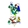
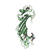
| Entry | Database: PDB / ID: 1ilr | ||||||
|---|---|---|---|---|---|---|---|
| Title | CRYSTAL STRUCTURE OF THE INTERLEUKIN-1 RECEPTOR ANTAGONIST | ||||||
 Components Components | INTERLEUKIN-1 RECEPTOR ANTAGONIST PROTEIN | ||||||
 Keywords Keywords | BINDING PROTEIN | ||||||
| Function / homology |  Function and homology information Function and homology informationinterleukin-1 type I receptor antagonist activity / interleukin-1 type II receptor antagonist activity / interleukin-1, type I receptor binding / interleukin-1 receptor antagonist activity / interleukin-1, type II receptor binding / negative regulation of interleukin-1-mediated signaling pathway / negative regulation of heterotypic cell-cell adhesion / interleukin-1 receptor binding / insulin secretion / Interleukin-10 signaling ...interleukin-1 type I receptor antagonist activity / interleukin-1 type II receptor antagonist activity / interleukin-1, type I receptor binding / interleukin-1 receptor antagonist activity / interleukin-1, type II receptor binding / negative regulation of interleukin-1-mediated signaling pathway / negative regulation of heterotypic cell-cell adhesion / interleukin-1 receptor binding / insulin secretion / Interleukin-10 signaling / response to glucocorticoid / cytokine activity / acute-phase response / lipid metabolic process / Interleukin-1 signaling / immune response / inflammatory response / centrosome / : / extracellular exosome / nucleoplasm / plasma membrane / cytosol Similarity search - Function | ||||||
| Biological species |  Homo sapiens (human) Homo sapiens (human) | ||||||
| Method |  X-RAY DIFFRACTION / Resolution: 2.1 Å X-RAY DIFFRACTION / Resolution: 2.1 Å | ||||||
 Authors Authors | Schreuder, H.A. / Rondeau, J.-M. / Tardif, C. | ||||||
 Citation Citation | Journal: Eur.J.Biochem. / Year: 1995 Title: Refined crystal structure of the interleukin-1 receptor antagonist. Presence of a disulfide link and a cis-proline. Authors: Schreuder, H.A. / Rondeau, J.M. / Tardif, C. / Soffientini, A. / Sarubbi, E. / Akeson, A. / Bowlin, T.L. / Yanofsky, S. / Barrett, R.W. | ||||||
| History |
|
- Structure visualization
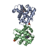
| Structure viewer | Molecule: MolmilJmol/JSmol |
|---|
- Downloads & links
-Download
| PDBx/mmCIF format | 1ilr.cif.gz | 69.7 KB | Display | PDBx/mmCIF format |
|---|---|---|---|---|
| PDB format | pdb1ilr.ent.gz | 52.3 KB | Display | PDB format |
| PDBx/mmJSON format | 1ilr.json.gz | Tree view | PDBx/mmJSON format | |
| Others |  Other downloads Other downloads |
-Validation report
| Arichive directory | https://data.pdbj.org/pub/pdb/validation_reports/il/1ilrftp://data.pdbj.org/pub/pdb/validation_reports/il/1ilr | HTTPS FTP |
|---|
-Related structure data
| Similar structure data |
|---|


-Links
 PDBj
PDBj


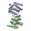
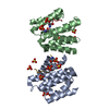
- Assembly
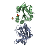
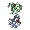
| Deposited unit | 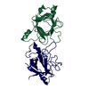
| ||||||||
|---|---|---|---|---|---|---|---|---|---|
| 1 |
| ||||||||
| Unit cell |
| ||||||||
| Atom site foot note | 1: CIS PROLINE - PRO 1 53 / 2: CIS PROLINE - PRO 2 53 | ||||||||
| Noncrystallographic symmetry (NCS) | NCS oper: (Code: given Matrix: (0.342973, 0.714236, -0.61011), Vector: |
-Components
| #1: Protein | Mass: 17145.406 Da / Num. of mol.: 2 Source method: isolated from a genetically manipulated source Source: (gene. exp.) Homo sapiens (human) / References: UniProt: P18510#2: Water | ChemComp-HOH / |  Mass: 18.015 Da / Num. of mol.: 139 / Source method: isolated from a natural source / Formula: H2O Mass: 18.015 Da / Num. of mol.: 139 / Source method: isolated from a natural source / Formula: H2OHas protein modification | Y | |
|---|
-Experimental details
-Experiment
| Experiment | Method: X-RAY DIFFRACTION |
|---|
- Sample preparation
| Crystal | Density Matthews: 2.17 Å3/Da / Density % sol: 43.39 % | ||||||||||||||||||||||||||||||||||||||||||||||||
|---|---|---|---|---|---|---|---|---|---|---|---|---|---|---|---|---|---|---|---|---|---|---|---|---|---|---|---|---|---|---|---|---|---|---|---|---|---|---|---|---|---|---|---|---|---|---|---|---|---|
| Crystal grow | *PLUS Temperature: 4 ℃ / Method: vapor diffusion, hanging drop | ||||||||||||||||||||||||||||||||||||||||||||||||
| Components of the solutions | *PLUS
|
-Data collection
| Radiation | Scattering type: x-ray |
|---|---|
| Radiation wavelength | Relative weight: 1 |
| Reflection | *PLUS Highest resolution: 2.1 Å / Num. obs: 16972 / % possible obs: 92.7 % |
| Reflection shell | *PLUS Highest resolution: 2.1 Å / Lowest resolution: 2.2 Å / % possible obs: 60.1 % |
- Processing
| Software |
| ||||||||||||||||||||||||||||||||||||||||||||||||||||||||||||
|---|---|---|---|---|---|---|---|---|---|---|---|---|---|---|---|---|---|---|---|---|---|---|---|---|---|---|---|---|---|---|---|---|---|---|---|---|---|---|---|---|---|---|---|---|---|---|---|---|---|---|---|---|---|---|---|---|---|---|---|---|---|
| Refinement | Resolution: 2.1→8 Å / σ(F): 0 /
| ||||||||||||||||||||||||||||||||||||||||||||||||||||||||||||
| Refinement step | Cycle: LAST / Resolution: 2.1→8 Å
| ||||||||||||||||||||||||||||||||||||||||||||||||||||||||||||
| Refine LS restraints |
| ||||||||||||||||||||||||||||||||||||||||||||||||||||||||||||
| Software | *PLUS Name: X-PLOR / Classification: refinement | ||||||||||||||||||||||||||||||||||||||||||||||||||||||||||||
| Refinement | *PLUS Rfactor obs: 0.197 / Rfactor Rwork: 0.197 | ||||||||||||||||||||||||||||||||||||||||||||||||||||||||||||
| Solvent computation | *PLUS | ||||||||||||||||||||||||||||||||||||||||||||||||||||||||||||
| Displacement parameters | *PLUS | ||||||||||||||||||||||||||||||||||||||||||||||||||||||||||||
| Refine LS restraints | *PLUS Type: x_angle_d / Dev ideal: 1.5 |
