 Movie
Movie Controller


+ Open data

- Basic information
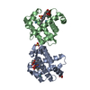
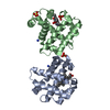
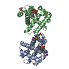
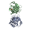
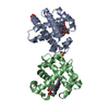
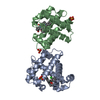
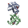
| Entry | Database: PDB / ID: 3lb2 | ||||||
|---|---|---|---|---|---|---|---|
| Title | Two-site competitive inhibition in dehaloperoxidase-hemoglobin | ||||||
 Components Components | DEHALOPEROXIDASE A | ||||||
 Keywords Keywords | OXIDOREDUCTASE / PEROXIDASE / GLOBIN | ||||||
| Function / homology |  Function and homology information Function and homology informationoxygen carrier activity / peroxidase activity / oxygen binding / heme binding / metal ion binding Similarity search - Function | ||||||
| Biological species |   AMPHITRITE ORNATA (invertebrata) AMPHITRITE ORNATA (invertebrata) | ||||||
| Method |  X-RAY DIFFRACTION / SYNCHROTRON / MOLECULAR REPLACEMENT / Resolution: 1.06 Å X-RAY DIFFRACTION / SYNCHROTRON / MOLECULAR REPLACEMENT / Resolution: 1.06 Å | ||||||
 Authors Authors | de Serrano, V.S. / Franzen, S. / Thompson, M.K. / Davis, M.F. / Nicoletti, F.P. / Howes, B.D. / Smulevich, G. | ||||||
 Citation Citation | Journal: Biophys.J. / Year: 2010 Title: Internal binding of halogenated phenols in dehaloperoxidase-hemoglobin inhibits peroxidase function. Authors: Thompson, M.K. / Davis, M.F. / de Serrano, V. / Nicoletti, F.P. / Howes, B.D. / Smulevich, G. / Franzen, S. | ||||||
| History |
|
- Structure visualization
| Structure viewer | Molecule: MolmilJmol/JSmol |
|---|
- Downloads & links
-Download
| PDBx/mmCIF format | 3lb2.cif.gz | 174.1 KB | Display | PDBx/mmCIF format |
|---|---|---|---|---|
| PDB format | pdb3lb2.ent.gz | 141.4 KB | Display | PDB format |
| PDBx/mmJSON format | 3lb2.json.gz | Tree view | PDBx/mmJSON format | |
| Others |  Other downloads Other downloads |
-Validation report
| Arichive directory | https://data.pdbj.org/pub/pdb/validation_reports/lb/3lb2ftp://data.pdbj.org/pub/pdb/validation_reports/lb/3lb2 | HTTPS FTP |
|---|
-Related structure data
| Related structure data |  3lb1C  3lb3C  3lb4C 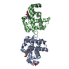 2qfkS C: citing same article ( S: Starting model for refinement |
|---|---|
| Similar structure data |


-Links
 PDBj
PDBj


- Assembly
| Deposited unit | 
| ||||||||
|---|---|---|---|---|---|---|---|---|---|
| 1 |
| ||||||||
| 2 | 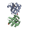
| ||||||||
| Unit cell |
|
-Components
| #1: Protein | Mass: 15548.597 Da / Num. of mol.: 2 Source method: isolated from a genetically manipulated source Source: (gene. exp.) AMPHITRITE ORNATA (invertebrata) / Gene: DHPA / Plasmid: PET16B / Production host:  #2: Chemical |   Mass: 616.487 Da / Num. of mol.: 2 / Source method: obtained synthetically / Formula: C34H32FeN4O4 Mass: 616.487 Da / Num. of mol.: 2 / Source method: obtained synthetically / Formula: C34H32FeN4O4#3: Chemical |   Mass: 173.007 Da / Num. of mol.: 2 / Source method: obtained synthetically / Formula: C6H5BrO Mass: 173.007 Da / Num. of mol.: 2 / Source method: obtained synthetically / Formula: C6H5BrO#4: Chemical |   Mass: 96.063 Da / Num. of mol.: 2 / Source method: obtained synthetically / Formula: SO4 Mass: 96.063 Da / Num. of mol.: 2 / Source method: obtained synthetically / Formula: SO4#5: Water | ChemComp-HOH / |  Mass: 18.015 Da / Num. of mol.: 356 / Source method: isolated from a natural source / Formula: H2O Mass: 18.015 Da / Num. of mol.: 356 / Source method: isolated from a natural source / Formula: H2O |
|---|
-Experimental details
-Experiment
| Experiment | Method: X-RAY DIFFRACTION / Number of used crystals: 1 |
|---|
- Sample preparation
| Crystal | Density Matthews: 2.18 Å3/Da / Density % sol: 43.59 % |
|---|---|
| Crystal grow | Temperature: 277 K / Method: vapor diffusion, hanging drop / pH: 5.9 Details: 0.2 M AMMONIUM SULFATE, 32% PEG 4000, PH 5.9, VAPOR DIFFUSION, HANGING DROP, TEMPERATURE 277K |
-Data collection
| Diffraction | Mean temperature: 100 K |
|---|---|
| Diffraction source | Source: SYNCHROTRON / Site: APS  / Beamline: 22-ID / Wavelength: 0.91942 / Wavelength: 0.91942 Å / Beamline: 22-ID / Wavelength: 0.91942 / Wavelength: 0.91942 Å |
| Detector | Type: MARMOSAIC 300 mm CCD / Detector: CCD / Date: Nov 11, 2007 |
| Radiation | Protocol: SINGLE WAVELENGTH / Monochromatic (M) / Laue (L): M / Scattering type: x-ray |
| Radiation wavelength | Wavelength: 0.91942 Å / Relative weight: 1 |
| Reflection | Resolution: 1.06→35 Å / Num. all: 121970 / Num. obs: 115854 / % possible obs: 98.7 % / Observed criterion σ(I): 2 / Redundancy: 4 % / Biso Wilson estimate: 6.8 Å2 / Rmerge(I) obs: 0.052 / Net I/σ(I): 22.5 |
| Reflection shell | Resolution: 1.06→1.09 Å / Redundancy: 3.3 % / Rmerge(I) obs: 0.318 / Mean I/σ(I) obs: 3.8 / Num. unique all: 8490 / % possible all: 93.8 |
- Processing
| Software |
| |||||||||||||||||||||||||||||||||||||||||||||||||||||||||||||||||||||||||||||||||||||||||||||||||||||||||
|---|---|---|---|---|---|---|---|---|---|---|---|---|---|---|---|---|---|---|---|---|---|---|---|---|---|---|---|---|---|---|---|---|---|---|---|---|---|---|---|---|---|---|---|---|---|---|---|---|---|---|---|---|---|---|---|---|---|---|---|---|---|---|---|---|---|---|---|---|---|---|---|---|---|---|---|---|---|---|---|---|---|---|---|---|---|---|---|---|---|---|---|---|---|---|---|---|---|---|---|---|---|---|---|---|---|---|
| Refinement | Method to determine structure: MOLECULAR REPLACEMENT Starting model: PDB ENTRY 2QFK Resolution: 1.06→35 Å / Cor.coef. Fo:Fc: 0.961 / Cor.coef. Fo:Fc free: 0.954 / SU B: 0.846 / SU ML: 0.02 / Cross valid method: THROUGHOUT / σ(I): 2 / ESU R: 0.038 / ESU R Free: 0.035 / Stereochemistry target values: MAXIMUM LIKELIHOOD / Details: HYDROGENS HAVE BEEN ADDED IN THE RIDING POSITIONS
| |||||||||||||||||||||||||||||||||||||||||||||||||||||||||||||||||||||||||||||||||||||||||||||||||||||||||
| Solvent computation | Ion probe radii: 0.8 Å / Shrinkage radii: 0.8 Å / VDW probe radii: 1.2 Å / Solvent model: MASK | |||||||||||||||||||||||||||||||||||||||||||||||||||||||||||||||||||||||||||||||||||||||||||||||||||||||||
| Displacement parameters | Biso mean: 10.17 Å2
| |||||||||||||||||||||||||||||||||||||||||||||||||||||||||||||||||||||||||||||||||||||||||||||||||||||||||
| Refinement step | Cycle: LAST / Resolution: 1.06→35 Å
| |||||||||||||||||||||||||||||||||||||||||||||||||||||||||||||||||||||||||||||||||||||||||||||||||||||||||
| Refine LS restraints |
| |||||||||||||||||||||||||||||||||||||||||||||||||||||||||||||||||||||||||||||||||||||||||||||||||||||||||
| LS refinement shell | Resolution: 1.06→1.09 Å / Total num. of bins used: 20
|
