 Movie
Movie Controller

[English] 日本語
 Yorodumi
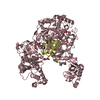
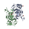
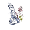
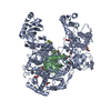
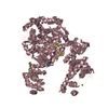
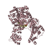
- PDB-1iq8: Crystal Structure of archaeosine tRNA-guanine transglycosylase fr... -
+ Open data

- Basic information
| Entry | Database: PDB / ID: 1iq8 | ||||||
|---|---|---|---|---|---|---|---|
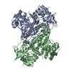
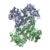
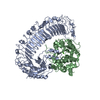
| Title | Crystal Structure of archaeosine tRNA-guanine transglycosylase from Pyrococcus horikoshii | ||||||
 Components Components | ARCHAEOSINE TRNA-GUANINE TRANSGLYCOSYLASE | ||||||
 Keywords Keywords | TRANSFERASE / (ALPHA/BETA)8 BARREL / RIKEN Structural Genomics/Proteomics Initiative / RSGI / Structural Genomics | ||||||
| Function / homology |  Function and homology information Function and homology informationtRNA-guanine15 transglycosylase / pentosyltransferase activity / tRNA wobble guanine modification / RNA binding / zinc ion binding / cytoplasm Similarity search - Function | ||||||
| Biological species |   Pyrococcus horikoshii (archaea) Pyrococcus horikoshii (archaea) | ||||||
| Method |  X-RAY DIFFRACTION / SYNCHROTRON / MAD / Resolution: 2.2 Å X-RAY DIFFRACTION / SYNCHROTRON / MAD / Resolution: 2.2 Å | ||||||
 Authors Authors | Ishitani, R. / Nureki, O. / Fukai, S. / Kijimoto, T. / Nameki, N. / Watanabe, M. / Kondo, H. / Sekine, M. / Okada, N. / Nishimura, S. ...Ishitani, R. / Nureki, O. / Fukai, S. / Kijimoto, T. / Nameki, N. / Watanabe, M. / Kondo, H. / Sekine, M. / Okada, N. / Nishimura, S. / Yokoyama, S. / RIKEN Structural Genomics/Proteomics Initiative (RSGI) | ||||||
 Citation Citation | Journal: J.Mol.Biol. / Year: 2002 Title: Crystal structure of archaeosine tRNA-guanine transglycosylase. Authors: Ishitani, R. / Nureki, O. / Fukai, S. / Kijimoto, T. / Nameki, N. / Watanabe, M. / Kondo, H. / Sekine, M. / Okada, N. / Nishimura, S. / Yokoyama, S. | ||||||
| History |
|
- Structure visualization
| Structure viewer | Molecule: MolmilJmol/JSmol |
|---|
- Downloads & links
-Download
| PDBx/mmCIF format | 1iq8.cif.gz | 240.3 KB | Display | PDBx/mmCIF format |
|---|---|---|---|---|
| PDB format | pdb1iq8.ent.gz | 194.5 KB | Display | PDB format |
| PDBx/mmJSON format | 1iq8.json.gz | Tree view | PDBx/mmJSON format | |
| Others |  Other downloads Other downloads |
-Validation report
| Arichive directory | https://data.pdbj.org/pub/pdb/validation_reports/iq/1iq8ftp://data.pdbj.org/pub/pdb/validation_reports/iq/1iq8 | HTTPS FTP |
|---|
-Related structure data


-Links
 PDBj
PDBj
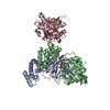
- Assembly
| Deposited unit | 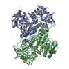
| ||||||||
|---|---|---|---|---|---|---|---|---|---|
| 1 |
| ||||||||
| Unit cell |
|
-Components
| #1: Protein | Mass: 66700.141 Da / Num. of mol.: 2 Source method: isolated from a genetically manipulated source Source: (gene. exp.) Pyrococcus horikoshii (archaea) / Plasmid: pET3A / Species (production host): Escherichia coli / Production host:  References: UniProt: O58843, tRNA-guanosine34 preQ1 transglycosylase #2: Chemical |   Mass: 65.409 Da / Num. of mol.: 2 / Source method: obtained synthetically / Formula: Zn Mass: 65.409 Da / Num. of mol.: 2 / Source method: obtained synthetically / Formula: Zn#3: Chemical |   Mass: 24.305 Da / Num. of mol.: 2 / Source method: obtained synthetically / Formula: Mg Mass: 24.305 Da / Num. of mol.: 2 / Source method: obtained synthetically / Formula: Mg#4: Water | ChemComp-HOH / |  Mass: 18.015 Da / Num. of mol.: 291 / Source method: isolated from a natural source / Formula: H2O Mass: 18.015 Da / Num. of mol.: 291 / Source method: isolated from a natural source / Formula: H2O |
|---|
-Experimental details
-Experiment
| Experiment | Method: X-RAY DIFFRACTION / Number of used crystals: 1 |
|---|
- Sample preparation
| Crystal | Density Matthews: 3.36 Å3/Da / Density % sol: 63.37 % |
|---|---|
| Crystal grow | Temperature: 293 K / Method: vapor diffusion, hanging drop / pH: 7.5 Details: sodium phosphate, potassium phosphate, sodium acetate, HEPES, pH 7.5, VAPOR DIFFUSION, HANGING DROP, temperature 293K |
| Crystal grow | *PLUS Details: Ishitani, R., (2001) Acta Cryst., D57, 1659. |
-Data collection
| Diffraction | Mean temperature: 100 K |
|---|---|
| Diffraction source | Source: SYNCHROTRON / Site: SPring-8  / Beamline: BL44XU / Wavelength: 0.9 Å / Beamline: BL44XU / Wavelength: 0.9 Å |
| Detector | Type: OXFORD PX210 / Detector: CCD / Date: Nov 20, 2000 / Details: mirrors |
| Radiation | Monochromator: Si 111 / Protocol: SINGLE WAVELENGTH / Monochromatic (M) / Laue (L): M / Scattering type: x-ray |
| Radiation wavelength | Wavelength: 0.9 Å / Relative weight: 1 |
| Reflection | Resolution: 2.2→50 Å / Num. all: 530165 / Num. obs: 530165 / % possible obs: 95.3 % / Observed criterion σ(F): 0 / Observed criterion σ(I): 0 / Redundancy: 6 % / Biso Wilson estimate: 19.7 Å2 / Rmerge(I) obs: 0.092 / Rsym value: 0.092 / Net I/σ(I): 34 |
| Reflection shell | Resolution: 2.2→2.24 Å / Redundancy: 4.1 % / Rmerge(I) obs: 0.256 / Mean I/σ(I) obs: 7.1 / Num. unique all: 4241 / Rsym value: 0.256 / % possible all: 93 |
| Reflection | *PLUS Lowest resolution: 50 Å / Num. obs: 88967 / Num. measured all: 530165 / Rmerge(I) obs: 0.092 |
| Reflection shell | *PLUS % possible obs: 93 % / Num. unique obs: 4241 / Num. measured obs: 17411 / Rmerge(I) obs: 0.256 / Mean I/σ(I) obs: 7.1 |
- Processing
| Software |
| ||||||||||||||||||||
|---|---|---|---|---|---|---|---|---|---|---|---|---|---|---|---|---|---|---|---|---|---|
| Refinement | Method to determine structure: MAD / Resolution: 2.2→50 Å / Isotropic thermal model: Overall / Cross valid method: THROUGHOUT / σ(F): 0 / σ(I): 0 / Stereochemistry target values: Engh & Huber
| ||||||||||||||||||||
| Displacement parameters | Biso mean: 40.4 Å2
| ||||||||||||||||||||
| Refinement step | Cycle: LAST / Resolution: 2.2→50 Å
| ||||||||||||||||||||
| Refinement | *PLUS Lowest resolution: 50 Å / Rfactor obs: 0.227 / Rfactor Rfree: 0.261 / Rfactor Rwork: 0.227 | ||||||||||||||||||||
| Solvent computation | *PLUS | ||||||||||||||||||||
| Displacement parameters | *PLUS | ||||||||||||||||||||
| Refine LS restraints | *PLUS
|
