 Movie
Movie Controller

[English] 日本語
 Yorodumi
- PDB-4ii3: Crystal structure of S. pombe Ubiquitin activating enzyme 1 (Uba1... -
+ Open data

- Basic information
| Entry | Database: PDB / ID: 4ii3 | ||||||
|---|---|---|---|---|---|---|---|
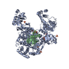
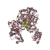
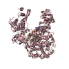
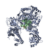
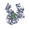
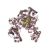
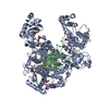
| Title | Crystal structure of S. pombe Ubiquitin activating enzyme 1 (Uba1) in complex with ubiquitin and ATP/Mg | ||||||
 Components Components |
| ||||||
 Keywords Keywords | LIGASE / Rossmann-like Fold / ubiquitin-like fold / Ubiquitin activating enzyme activity / ATP binding / Ligase activity / ATP/Mg binding / Ubiquitin E2 binding | ||||||
| Function / homology |  Function and homology information Function and homology information: / Antigen processing: Ubiquitination & Proteasome degradation / E1 ubiquitin-activating enzyme / ubiquitin activating enzyme activity / ribosomal large subunit export from nucleus / modification-dependent protein catabolic process / protein tag activity / cytosolic large ribosomal subunit / ubiquitin-dependent protein catabolic process / cytoplasmic translation ...: / Antigen processing: Ubiquitination & Proteasome degradation / E1 ubiquitin-activating enzyme / ubiquitin activating enzyme activity / ribosomal large subunit export from nucleus / modification-dependent protein catabolic process / protein tag activity / cytosolic large ribosomal subunit / ubiquitin-dependent protein catabolic process / cytoplasmic translation / structural constituent of ribosome / protein ubiquitination / DNA damage response / ubiquitin protein ligase binding / nucleolus / magnesium ion binding / ATP hydrolysis activity / ATP binding / nucleus / cytoplasm / cytosol Similarity search - Function | ||||||
| Biological species |  | ||||||
| Method |  X-RAY DIFFRACTION / SYNCHROTRON / MOLECULAR REPLACEMENT / Resolution: 2.9 Å X-RAY DIFFRACTION / SYNCHROTRON / MOLECULAR REPLACEMENT / Resolution: 2.9 Å | ||||||
 Authors Authors | Olsen, S.K. / Lima, C.D. | ||||||
 Citation Citation | Journal: Mol.Cell / Year: 2013 Title: Structure of a ubiquitin E1-E2 complex: insights to E1-E2 thioester transfer. Authors: Olsen, S.K. / Lima, C.D. | ||||||
| History |
|
- Structure visualization
| Structure viewer | Molecule: MolmilJmol/JSmol |
|---|
- Downloads & links
-Download
| PDBx/mmCIF format | 4ii3.cif.gz | 434.7 KB | Display | PDBx/mmCIF format |
|---|---|---|---|---|
| PDB format | pdb4ii3.ent.gz | 342.2 KB | Display | PDB format |
| PDBx/mmJSON format | 4ii3.json.gz | Tree view | PDBx/mmJSON format | |
| Others |  Other downloads Other downloads |
-Validation report
| Arichive directory | https://data.pdbj.org/pub/pdb/validation_reports/ii/4ii3ftp://data.pdbj.org/pub/pdb/validation_reports/ii/4ii3 | HTTPS FTP |
|---|
-Related structure data
| Related structure data |  4ii2C  3cmmS C: citing same article ( S: Starting model for refinement |
|---|---|
| Similar structure data |


-Links
 PDBj
PDBj


- Assembly
| Deposited unit | 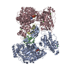
| ||||||||
|---|---|---|---|---|---|---|---|---|---|
| 1 | 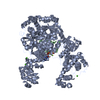
| ||||||||
| 2 | 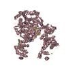
| ||||||||
| Unit cell |
|
-Components
-Protein , 2 types, 4 molecules ACBD
| #1: Protein | Mass: 111764.047 Da / Num. of mol.: 2 / Fragment: Uba1, UNP residues 13-1012 Source method: isolated from a genetically manipulated source Details: N-terminal SMT3 fusion (Ulp-cleavable) Source: (gene. exp.) Strain: 972 / ATCC 24843 Gene: ptr3, SPBC1604.21c, SPBC211.09, Ubiquitin activating enzyme 1 (Uba1) Plasmid: pSMT3 / Production host:  #2: Protein | Mass: 10740.132 Da / Num. of mol.: 2 / Fragment: UNP residues 1-76 Source method: isolated from a genetically manipulated source Source: (gene. exp.) Strain: strain 972 / ATCC 24843 / Gene: ubi2 / Plasmid: pET-28 / Production host: |
|---|
-Non-polymers , 4 types, 165 molecules 


| #3: Chemical | ChemComp-MG /  Mass: 24.305 Da / Num. of mol.: 4 / Source method: obtained synthetically / Formula: Mg Mass: 24.305 Da / Num. of mol.: 4 / Source method: obtained synthetically / Formula: Mg#4: Chemical |  Mass: 507.181 Da / Num. of mol.: 2 / Source method: obtained synthetically / Formula: C10H16N5O13P3 / Comment: ATP, energy-carrying molecule*YM Mass: 507.181 Da / Num. of mol.: 2 / Source method: obtained synthetically / Formula: C10H16N5O13P3 / Comment: ATP, energy-carrying molecule*YM#5: Chemical | ChemComp-CA /  Mass: 40.078 Da / Num. of mol.: 10 / Source method: obtained synthetically / Formula: Ca Mass: 40.078 Da / Num. of mol.: 10 / Source method: obtained synthetically / Formula: Ca#6: Water | ChemComp-HOH / | Mass: 18.015 Da / Num. of mol.: 149 / Source method: isolated from a natural source / Formula: H2O |
|---|
-Experimental details
-Experiment
| Experiment | Method: X-RAY DIFFRACTION / Number of used crystals: 1 |
|---|
- Sample preparation
| Crystal | Density Matthews: 2.64 Å3/Da / Density % sol: 53.44 % |
|---|---|
| Crystal grow | Temperature: 291 K / Method: vapor diffusion, hanging drop / pH: 6.5 Details: 0.1M Sodium Cacodylate, 0.2M Calcium Acetate, 11.5% Peg8000, 3% 1,5 diaminopentane, pH 6.5, VAPOR DIFFUSION, HANGING DROP, temperature 291K |
-Data collection
| Diffraction | Mean temperature: 108 K | |||||||||||||||||||||||||||||||||||||||||||||||||||||||||||||||||||||||||||||
|---|---|---|---|---|---|---|---|---|---|---|---|---|---|---|---|---|---|---|---|---|---|---|---|---|---|---|---|---|---|---|---|---|---|---|---|---|---|---|---|---|---|---|---|---|---|---|---|---|---|---|---|---|---|---|---|---|---|---|---|---|---|---|---|---|---|---|---|---|---|---|---|---|---|---|---|---|---|---|
| Diffraction source | Source: SYNCHROTRON / Site: APS  / Beamline: 24-ID-E / Wavelength: 0.979 Å / Beamline: 24-ID-E / Wavelength: 0.979 Å | |||||||||||||||||||||||||||||||||||||||||||||||||||||||||||||||||||||||||||||
| Detector | Type: ADSC QUANTUM 315 / Detector: CCD / Date: Apr 16, 2012 | |||||||||||||||||||||||||||||||||||||||||||||||||||||||||||||||||||||||||||||
| Radiation | Monochromator: Si(111) / Protocol: SINGLE WAVELENGTH / Monochromatic (M) / Laue (L): M / Scattering type: x-ray | |||||||||||||||||||||||||||||||||||||||||||||||||||||||||||||||||||||||||||||
| Radiation wavelength | Wavelength: 0.979 Å / Relative weight: 1 | |||||||||||||||||||||||||||||||||||||||||||||||||||||||||||||||||||||||||||||
| Reflection | Redundancy: 3.4 % / Av σ(I) over netI: 8 / Number: 189068 / Rmerge(I) obs: 0.113 / Χ2: 1.07 / D res high: 2.9 Å / D res low: 50 Å / Num. obs: 55035 / % possible obs: 95.2 | |||||||||||||||||||||||||||||||||||||||||||||||||||||||||||||||||||||||||||||
| Diffraction reflection shell |
| |||||||||||||||||||||||||||||||||||||||||||||||||||||||||||||||||||||||||||||
| Reflection | Resolution: 2.9→50 Å / Num. all: 57810 / Num. obs: 55035 / % possible obs: 95.2 % / Observed criterion σ(I): 0 / Redundancy: 3.4 % / Rmerge(I) obs: 0.113 / Χ2: 1.07 / Net I/σ(I): 8.1 | |||||||||||||||||||||||||||||||||||||||||||||||||||||||||||||||||||||||||||||
| Reflection shell |
|
- Processing
| Software |
| |||||||||||||||||||||||||||||||||||||||||||||||||||||||||||||||||||||||||||||
|---|---|---|---|---|---|---|---|---|---|---|---|---|---|---|---|---|---|---|---|---|---|---|---|---|---|---|---|---|---|---|---|---|---|---|---|---|---|---|---|---|---|---|---|---|---|---|---|---|---|---|---|---|---|---|---|---|---|---|---|---|---|---|---|---|---|---|---|---|---|---|---|---|---|---|---|---|---|---|
| Refinement | Method to determine structure: MOLECULAR REPLACEMENT Starting model: PDB ENTRY 3CMM Resolution: 2.9→50 Å / Occupancy max: 1 / Occupancy min: 1 / σ(F): 0 / Stereochemistry target values: Engh & Huber
| |||||||||||||||||||||||||||||||||||||||||||||||||||||||||||||||||||||||||||||
| Solvent computation | Bsol: 11.0687 Å2 | |||||||||||||||||||||||||||||||||||||||||||||||||||||||||||||||||||||||||||||
| Displacement parameters | Biso max: 136.35 Å2 / Biso mean: 61.8011 Å2 / Biso min: 2.79 Å2
| |||||||||||||||||||||||||||||||||||||||||||||||||||||||||||||||||||||||||||||
| Refinement step | Cycle: LAST / Resolution: 2.9→50 Å
| |||||||||||||||||||||||||||||||||||||||||||||||||||||||||||||||||||||||||||||
| Refine LS restraints |
| |||||||||||||||||||||||||||||||||||||||||||||||||||||||||||||||||||||||||||||
| LS refinement shell | Refine-ID: X-RAY DIFFRACTION / Total num. of bins used: 10
| |||||||||||||||||||||||||||||||||||||||||||||||||||||||||||||||||||||||||||||
| Xplor file |
|
