 ムービー
ムービー コントローラー
 構造ビューア
 EMN検索について


-検索条件
-検索結果
検索 (著者・登録者: da & fonseca & p)の結果全47件を表示しています
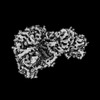
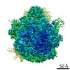
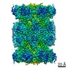
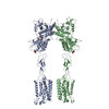
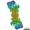
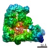
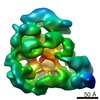
EMDB-40751: 
Isobutyryl-CoA mutase fused Q341A in the presence of GTP
PDB-8ssl:
Isobutyryl-CoA mutase fused Q341A in the presence of GTP

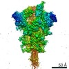
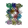
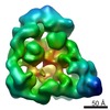
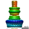
EMDB-40758:
Isobutyryl-CoA mutase fused in the presence of GMPPCP

PDB-8sta:
Isobutyryl-CoA mutase fused in the presence of GMPPCP
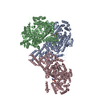
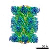
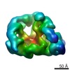
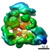
EMDB-23355:
Structure of dNTPase at 3.3 Angstrom Resolution
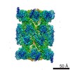
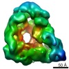
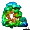
EMDB-23356:
Structure of dNTPase at 3.6 Angstrom Resolution

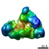
EMDB-23357:
Structure of 80S ribosome from EMPIAR-10064 at 5.6 Angstrom Resolution
EMDB-23358:
Structure of E. coli 70S ribosome from EMPIAR-10304 at 4.8 Angstrom Resolution

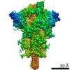
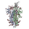
EMDB-11328:
SARS-CoV-2 spike in prefusion state
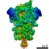
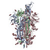
EMDB-11336:
SARS-CoV-2 spike in prefusion state (flexibility analysis, 1-up closed conformation)
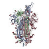
EMDB-11337:
SARS-CoV-2 spike in prefusion state (flexibility analysis, 1-up open conformation)
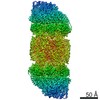
EMDB-11341:
SARS-CoV-2 stabilized spike in prefusion state (1-up conformation)
PDB-6zow:
SARS-CoV-2 spike in prefusion state
PDB-6zp5:
SARS-CoV-2 spike in prefusion state (flexibility analysis, 1-up closed conformation)
PDB-6zp7:
SARS-CoV-2 spike in prefusion state (flexibility analysis, 1-up open conformation)
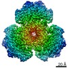
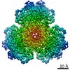
EMDB-4860:
Human 20S-PA200 Proteasome Complex
EMDB-4877:
Human 20S Proteasome

PDB-6rey:
Human 20S-PA200 Proteasome Complex

PDB-6rgq:
Human 20S Proteasome
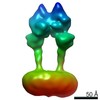
EMDB-0347:
Apo form metabotropic glutamate receptor 5 with Nanobody 43

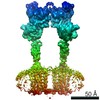
EMDB-0345:
Metabotropic Glutamate Receptor 5 bound to L-quisqualate and Nb43
EMDB-0346:
Metabotropic Glutamate Receptor 5 Apo Form
PDB-6n51:
Metabotropic Glutamate Receptor 5 bound to L-quisqualate and Nb43
PDB-6n52:
Metabotropic Glutamate Receptor 5 Apo Form
PDB-5fmg:
Structure and function based design of Plasmodium-selective proteasome inhibitors
EMDB-3231:
Structure and function based design of Plasmodium-selective proteasome inhibitors

PDB-5a0q:
Cryo-EM reveals the conformation of a substrate analogue in the human 20S proteasome core
EMDB-2981:
Cryo-EM reveals the conformation of a substrate analogue in the human 20S proteasome core

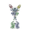
EMDB-2226:
Single particle analysis of recombinant human anaphase promoting complex (APC/C)
EMDB-2173:
Negative stain electron microscopy of a CSN-SCF~Nedd8/Skp2/Cks1 complex
EMDB-2174:
Negative stain electron microscopy of a CSN-SCF~Nedd8/Fbw7 complex

EMDB-2175:
Negative stain electron microscopy of a CSN(dCsn5)-SCF~Nedd8/Skp2/Cks1 complex
EMDB-2176:
Negative stain electron microscopy of a CSN complex
EMDB-2177:
Negative stain electron microscopy of a CSN complex
EMDB-2047:
Structure of the human 26S proteasome

EMDB-2026:
Structure of the proteasome subunit Rpn1
EMDB-1842:
Structural basis for the subunit assembly of the anaphase promoting complex

EMDB-1843:
Structural basis for the subunit assembly of the anaphase promoting complex

EMDB-1841:
Structural basis for the subunit assembly of the anaphase promoting complex
EMDB-1844:
Structural basis for the subunit assembly of the anaphase promoting complex
EMDB-1845:
Structural basis for the subunit assembly of the anaphase promoting complex
EMDB-1815:
The structure of anaphase promoting complex-Cdh1-Dbox at 10 Angstroms resolution
EMDB-1817:
Structure of S. cerevisiae anaphase promoting complex with the co-activator Cdh1
EMDB-1818:
Structure of S. cerevisiae anaphase promoting complex-Cdh1-KENbox peptide
EMDB-1816:
Structure of S. cerevisiae anaphase promoting complex
EMDB-1819:
Structure of S. cerevisiae anaphase promoting complex-Cdh1 bound to a Hsl1 fragment
EMDB-1617:
C12 symmetrised 3D reconstruction of the Shigella flexneri T3SS needle complex from negatively stained sample electron micrographs
 wwPDBはEMDBデータモデルのバージョン3へ移行します
