 Movie
Movie Controller

[English] 日本語
 Yorodumi
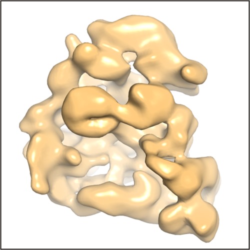
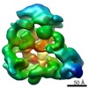
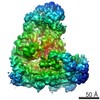
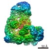
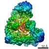
- EMDB-1819: Structure of S. cerevisiae anaphase promoting complex-Cdh1 bound ... -
+ Open data

- Basic information
| Entry | Database: EMDB / ID: EMD-1819 | |||||||||
|---|---|---|---|---|---|---|---|---|---|---|
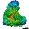
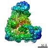
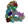
| Title | Structure of S. cerevisiae anaphase promoting complex-Cdh1 bound to a Hsl1 fragment | |||||||||
 Map data Map data | Structure of S. cerevisiae anaphase promoting complex-Cdh1 bound to a Hsl1 fragment | |||||||||
 Sample Sample |
| |||||||||
 Keywords Keywords | anaphase promoting complex / cyclosome / APC / APC/C / cell cycle / D-box / KEN-box / co-activator / Cdh1 / tetraticopeptide repeats / TPR / ubiquitylation / cyclin | |||||||||
| Biological species |  | |||||||||
| Method | single particle reconstruction / negative staining | |||||||||
 Authors Authors | daFonseca PCA / Kong EH / Zhang Z / Schreiber A / Williams MA / Morris EP / Barford D | |||||||||
 Citation Citation | Journal: Nature / Year: 2011 Title: Structures of APC/C(Cdh1) with substrates identify Cdh1 and Apc10 as the D-box co-receptor. Authors: Paula C A da Fonseca / Eric H Kong / Ziguo Zhang / Anne Schreiber / Mark A Williams / Edward P Morris / David Barford /  Abstract: The ubiquitylation of cell-cycle regulatory proteins by the large multimeric anaphase-promoting complex (APC/C) controls sister chromatid segregation and the exit from mitosis. Selection of APC/C ...The ubiquitylation of cell-cycle regulatory proteins by the large multimeric anaphase-promoting complex (APC/C) controls sister chromatid segregation and the exit from mitosis. Selection of APC/C targets is achieved through recognition of destruction motifs, predominantly the destruction (D)-box and KEN (Lys-Glu-Asn)-box. Although this process is known to involve a co-activator protein (either Cdc20 or Cdh1) together with core APC/C subunits, the structural basis for substrate recognition and ubiquitylation is not understood. Here we investigate budding yeast APC/C using single-particle electron microscopy and determine a cryo-electron microscopy map of APC/C in complex with the Cdh1 co-activator protein (APC/C(Cdh1)) bound to a D-box peptide at ∼10 Å resolution. We find that a combined catalytic and substrate-recognition module is located within the central cavity of the APC/C assembled from Cdh1, Apc10--a core APC/C subunit previously implicated in substrate recognition--and the cullin domain of Apc2. Cdh1 and Apc10, identified from difference maps, create a co-receptor for the D-box following repositioning of Cdh1 towards Apc10. Using NMR spectroscopy we demonstrate specific D-box-Apc10 interactions, consistent with a role for Apc10 in directly contributing towards D-box recognition by the APC/C(Cdh1) complex. Our results rationalize the contribution of both co-activator and core APC/C subunits to D-box recognition and provide a structural framework for understanding mechanisms of substrate recognition and catalysis by the APC/C. | |||||||||
| History |
|
- Structure visualization
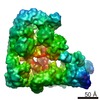
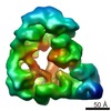
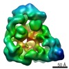
| Movie |
 Movie viewer Movie viewer |
|---|---|
| Structure viewer | EM map: SurfViewMolmilJmol/JSmol |
| Supplemental images |
- Downloads & links
-EMDB archive
| Map data | emd_1819.map.gz | 10 MB | EMDB map data format | |
|---|---|---|---|---|
| Header (meta data) | emd-1819-v30.xmlemd-1819.xml | 8.4 KB 8.4 KB | Display Display | EMDB header |
| Images | figure_EMD1819.tif | 208.4 KB | ||
| Archive directory |  http://ftp.pdbj.org/pub/emdb/structures/EMD-1819ftp://ftp.pdbj.org/pub/emdb/structures/EMD-1819 http://ftp.pdbj.org/pub/emdb/structures/EMD-1819ftp://ftp.pdbj.org/pub/emdb/structures/EMD-1819 | HTTPS FTP |
-Related structure data
-Links
| EMDB pages | EMDB (EBI/PDBe) / EMDataResource |
|---|
-Map
| File | Download / File: emd_1819.map.gz / Format: CCP4 / Size: 10.2 MB / Type: IMAGE STORED AS FLOATING POINT NUMBER (4 BYTES) | ||||||||||||||||||||||||||||||||||||||||||||||||||||||||||||||||||||
|---|---|---|---|---|---|---|---|---|---|---|---|---|---|---|---|---|---|---|---|---|---|---|---|---|---|---|---|---|---|---|---|---|---|---|---|---|---|---|---|---|---|---|---|---|---|---|---|---|---|---|---|---|---|---|---|---|---|---|---|---|---|---|---|---|---|---|---|---|---|
| Annotation | Structure of S. cerevisiae anaphase promoting complex-Cdh1 bound to a Hsl1 fragment | ||||||||||||||||||||||||||||||||||||||||||||||||||||||||||||||||||||
| Projections & slices | Image control
Images are generated by Spider. | ||||||||||||||||||||||||||||||||||||||||||||||||||||||||||||||||||||
| Voxel size | X=Y=Z: 3.47 Å | ||||||||||||||||||||||||||||||||||||||||||||||||||||||||||||||||||||
| Density |
| ||||||||||||||||||||||||||||||||||||||||||||||||||||||||||||||||||||
| Symmetry | Space group: 1 | ||||||||||||||||||||||||||||||||||||||||||||||||||||||||||||||||||||
| Details | EMDB XML:
CCP4 map header:
| ||||||||||||||||||||||||||||||||||||||||||||||||||||||||||||||||||||
 Y (Sec.)
 X (Row.)
 Z (Col.)


-Supplemental data
- Sample components
-Entire : S. cerevisiae anaphase promoting complex-Cdh1 bound to a Hsl1 fragment
| Entire | Name: S. cerevisiae anaphase promoting complex-Cdh1 bound to a Hsl1 fragment |
|---|---|
| Components |
|
-Supramolecule #1000: S. cerevisiae anaphase promoting complex-Cdh1 bound to a Hsl1 fragment
| Supramolecule | Name: S. cerevisiae anaphase promoting complex-Cdh1 bound to a Hsl1 fragment type: sample / ID: 1000 / Number unique components: 3 |
|---|
-Macromolecule #1: Anaphase promoting complex
| Macromolecule | Name: Anaphase promoting complex / type: protein_or_peptide / ID: 1 / Name.synonym: Anaphase promoting complex or cyclosome / Recombinant expression: No |
|---|---|
| Source (natural) | Organism: |
-Macromolecule #2: Cdh1
| Macromolecule | Name: Cdh1 / type: protein_or_peptide / ID: 2 / Name.synonym: Cdh1 / Recombinant expression: Yes |
|---|---|
| Source (natural) | Organism: |
| Recombinant expression | Organism:   Spodoptera frugiperda (fall armyworm) Spodoptera frugiperda (fall armyworm) |
-Macromolecule #3: Hsl1
| Macromolecule | Name: Hsl1 / type: protein_or_peptide / ID: 3 / Name.synonym: Hsl1 fragment / Recombinant expression: Yes |
|---|---|
| Source (natural) | Organism: |
| Recombinant expression | Organism:  |
-Experimental details
-Structure determination
| Method | negative staining |
|---|---|
 Processing Processing | single particle reconstruction |
| Aggregation state | particle |
-Sample preparation
| Staining | Type: NEGATIVE / Details: Grids were stained using 2% w/v uranyl acetate |
|---|---|
| Vitrification | Cryogen name: NONE / Instrument: OTHER |
- Electron microscopy
| Microscope | FEI TECNAI F20 |
|---|---|
| Details | Sample stained using uranyl acetate and imaged at room temperature |
| Image recording | Category: CCD / Film or detector model: TVIPS TEMCAM-F415 (4k x 4k) / Average electron dose: 100 e/Å2 |
| Electron beam | Acceleration voltage: 200 kV / Electron source:  FIELD EMISSION GUN FIELD EMISSION GUN |
| Electron optics | Illumination mode: FLOOD BEAM / Imaging mode: BRIGHT FIELD / Nominal magnification: 50000 |
| Sample stage | Specimen holder: Room temperature side entry / Specimen holder model: OTHER |
| Experimental equipment |  Model: Tecnai F20 / Image courtesy: FEI Company |
-Image processing
| Final reconstruction | Applied symmetry - Point group: C1 (asymmetric) |
|---|
