 Movie
Movie Controller
 Structure viewers
 About EMN search


-Search query
-Search result
Showing 1 - 50 of 30,364 items for (author: ma & c)

EMDB entry, No image
EMDB-43506: 
Cryo-EM structure of LKB1-STRADalpha-MO25alpha heterocomplex
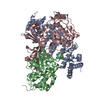
PDB-8vsu:
Cryo-EM structure of LKB1-STRADalpha-MO25alpha heterocomplex
EMDB entry, No image
EMDB-36836:
Cryo-EM structure of the human 55S mitoribosome with Tigecycline
EMDB entry, No image
EMDB-36837:
Cryo-EM structure of the human 39S mitoribosome with Tigecycline
EMDB entry, No image
EMDB-36838:
Cryo-EM structure of the human 80S ribosome with Tigecycline
EMDB entry, No image
EMDB-36839:
Cryo-EM structure of the yeast 80S ribosome with tigecycline, eEF2, Stm1 and eIF5A
EMDB entry, No image
EMDB-36945:
Cryo-EM structure of the yeast 80S ribosome with tigecycline, Not5 and P-site tRNA
EMDB entry, No image
EMDB-38629:
Cryo-EM structure of the human 80S ribosome with Tigecycline, E-tRNA, SERBP1 and eEF2
EMDB entry, No image
EMDB-38630:
Cryo-EM structure of the human 80S ribosome with Tigecycline, e-tRNA and CCDC124 (40S head Swivelled)
EMDB entry, No image
EMDB-38631:
Cryo-EM structure of the human 80S ribosome with Tigecycline, E-tRNA and P-tRNA
EMDB entry, No image
EMDB-38632:
Cryo-EM structure of the human 55S mitoribosome with 5um Tigecycline
EMDB entry, No image
EMDB-38633:
Cryo-EM structure of the human 39S mitoribosome with 5uM Tigecycline
EMDB entry, No image
EMDB-38634:
Cryo-EM structure of the human 55S mitoribosome with 10uM Tigecycline
EMDB entry, No image
EMDB-38635:
Cryo-EM structure of the human 39S mitoribosome with 10uM Tigecycline
EMDB entry, No image
EMDB-38636:
Cryo-EM structure of the human 55S mitoribosome with 5uM Tigecycline (focusing on 39S ribosome)
EMDB entry, No image
EMDB-38637:
Cryo-EM structure of the human 55S mitoribosome with 5uM Tigecycline (focusing on 28S ribosome head)
EMDB entry, No image
EMDB-38638:
Cryo-EM structure of the human 55S mitoribosome with 10uM Tigecycline (focusing on 39S ribosome)
EMDB entry, No image
EMDB-38639:
Cryo-EM structure of the human 55S mitoribosome with 10uM Tigecycline (focusing on 28S ribosome head)
EMDB entry, No image
EMDB-39455:
Cryo-EM structure of the human 80S ribosome with 100 um Tigecycline
EMDB entry, No image
EMDB-39456:
Cryo-EM structure of the human 80S ribosome with 4 um Tigecycline

PDB-8k2a:
Cryo-EM structure of the human 55S mitoribosome with Tigecycline

PDB-8k2b:
Cryo-EM structure of the human 39S mitoribosome with Tigecycline

PDB-8k2c:
Cryo-EM structure of the human 80S ribosome with Tigecycline

PDB-8k2d:
Cryo-EM structure of the yeast 80S ribosome with tigecycline, eEF2, Stm1 and eIF5A

PDB-8k82:
Cryo-EM structure of the yeast 80S ribosome with tigecycline, Not5 and P-site tRNA

PDB-8xsx:
Cryo-EM structure of the human 80S ribosome with Tigecycline, E-tRNA, SERBP1 and eEF2

PDB-8xsy:
Cryo-EM structure of the human 80S ribosome with Tigecycline, e-tRNA and CCDC124 (40S head Swivelled)

PDB-8xsz:
Cryo-EM structure of the human 80S ribosome with Tigecycline, E-tRNA and P-tRNA

PDB-8xt0:
Cryo-EM structure of the human 55S mitoribosome with 5um Tigecycline

PDB-8xt1:
Cryo-EM structure of the human 39S mitoribosome with 5uM Tigecycline

PDB-8xt2:
Cryo-EM structure of the human 55S mitoribosome with 10uM Tigecycline

PDB-8xt3:
Cryo-EM structure of the human 39S mitoribosome with 10uM Tigecycline

PDB-8yoo:
Cryo-EM structure of the human 80S ribosome with 100 um Tigecycline

PDB-8yop:
Cryo-EM structure of the human 80S ribosome with 4 um Tigecycline
EMDB entry, No image
EMDB-42979:
Engaged conformation of the human mitochondrial DNA polymerase gamma bound to DNA
EMDB entry, No image
EMDB-42980:
Human mitochondrial DNA polymerase gamma bound to a replication fork in an open conformation
EMDB entry, No image
EMDB-42982:
Human mitochondrial DNA polymerase catalytic subunit, PolG, in an APO conformation
EMDB entry, No image
EMDB-42984:
Active conformation of DNA polymerase gamma bound to DNA
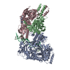
PDB-8v54:
Engaged conformation of the human mitochondrial DNA polymerase gamma bound to DNA
PDB-8v55:
Human mitochondrial DNA polymerase gamma bound to a replication fork in an open conformation
PDB-8v5d:
Human mitochondrial DNA polymerase catalytic subunit, PolG, in an APO conformation
PDB-8v5r:
Active conformation of DNA polymerase gamma bound to DNA
EMDB entry, No image
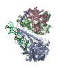
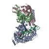
EMDB-50447:
Structure of the C. elegans Intron Lariat Spliceosome (Map 1)
EMDB entry, No image
EMDB-50449:
Structure of the C. elegans Intron Lariat Spliceosome (Map 2)
EMDB entry, No image
EMDB-50450:
Structure of the C. elegans Intron Lariat Spliceosome (Map 3)
EMDB entry, No image
EMDB-50451:
Structure of the C. elegans Intron Lariat Spliceosome (Map 4)
EMDB entry, No image
EMDB-50452:
Structure of the C. elegans Intron Lariat Spliceosome (Map 5)
EMDB entry, No image
EMDB-50453:
Structure of the C. elegans Intron Lariat Spliceosome (Map 6)
EMDB entry, No image
EMDB-50454:
Structure of the C. elegans Intron Lariat Spliceosome (Map 7)
EMDB entry, No image
EMDB-50455:
Structure of the C. elegans Intron Lariat Spliceosome (Map 8)
Pages:
 wwPDB to switch to version 3 of the EMDB data model
