 ムービー
ムービー コントローラー
 構造ビューア
 EMN検索について


-検索条件
-検索結果
検索 (著者・登録者: hui & s)の結果3,617件中、1から50件目までを表示しています

EMDBエントリ 画像なし
EMDB-38580: 
Structure of human class T GPCR TAS2R14-miniGs/gust complex with Aristolochic acid A.
EMDBエントリ 画像なし
EMDB-38582:
Structure of human class T GPCR TAS2R14-DNGi complex with Aristolochic acid A.
EMDBエントリ 画像なし
EMDB-38583:
Structure of human class T GPCR TAS2R14-Gi complex with Aristolochic acid A.
EMDBエントリ 画像なし
EMDB-38584:
Structure of human class T GPCR TAS2R14-Gustducin complex with Aristolochic acid A.
EMDBエントリ 画像なし
EMDB-38586:
Structure 2 of human class T GPCR TAS2R14-miniGs/gust complex with Flufenamic acid.
EMDBエントリ 画像なし
EMDB-38587:
Structure of human class T GPCR TAS2R14-DNGi complex with Flufenamic acid.
EMDBエントリ 画像なし
EMDB-38588:
Structure of human class T GPCR TAS2R14-Gi complex.
EMDBエントリ 画像なし
EMDB-39376:
Structure of human class T GPCR TAS2R14-Ggustducin complex with agonist 28.1
PDB-8xql:
Structure of human class T GPCR TAS2R14-miniGs/gust complex with Aristolochic acid A.
PDB-8xqn:
Structure of human class T GPCR TAS2R14-DNGi complex with Aristolochic acid A.
PDB-8xqo:
Structure of human class T GPCR TAS2R14-Gi complex with Aristolochic acid A.
PDB-8xqp:
Structure of human class T GPCR TAS2R14-Gustducin complex with Aristolochic acid A.
PDB-8xqr:
Structure 2 of human class T GPCR TAS2R14-miniGs/gust complex with Flufenamic acid.
PDB-8xqs:
Structure of human class T GPCR TAS2R14-DNGi complex with Flufenamic acid.
PDB-8xqt:
Structure of human class T GPCR TAS2R14-Gi complex.
PDB-8yky:
Structure of human class T GPCR TAS2R14-Ggustducin complex with agonist 28.1
EMDB-38216:
Cryo-EM structure of SARS-CoV-2 S-BQ.1 in complex with antibody O5C2
PDB-8xbf:
Cryo-EM structure of SARS-CoV-2 S-BQ.1 in complex with antibody O5C2

EMDB-36730:
SARS-CoV-2 Spike RBD (dimer) in complex with two 2S-1244 nanobodies
EMDB-36735:
Dimer of SARS-CoV-2 BA.2 spike and IBT-CoV144(C3 symmetry)
EMDB-36740:
Dimer of SARS-CoV-2 BA.2 spike and IBT-CoV144(C1 symmetry)

PDB-8jys:
SARS-CoV-2 Spike RBD (dimer) in complex with two 2S-1244 nanobodies

EMDB-38613:
Structure of MPXV B6 and D68 fab complex

PDB-8xs3:
Structure of MPXV B6 and D68 fab complex
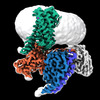
EMDB-37957:
Cryo-EM map for Mumps Virus L Protein Bound by Phosphoprotein Tetramer
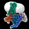
EMDB-37958:
Cryo-EM map for Mumps Virus L Protein Bound by Phosphoprotein Tetramer (Focused map for CD-MTase-CTD)
EMDB-37959:
Cryo-EM map for Mumps Virus L Protein Bound by Phosphoprotein Tetramer (Focused map for RdRp-PRNTase)
EMDB-37960:
Cryo-EM map for Mumps Virus L Protein Bound by Phosphoprotein Tetramer (Focused map for tetrameric phosphoproteins)
EMDB-37961:
Cryo-EM map for Mumps Virus L Protein (State 2) Bound by Phosphoprotein Tetramer
EMDB-37962:
Cryo-EM map for Mumps Virus L protein (state2) Bound by Phosphoprotein Tetramer (Focused for tetrameric phosphoprotein)
EMDB-37964:
Structure of the Mumps Virus L Protein (state2) Bound by Phosphoprotein Tetramer (composite map)

PDB-8x01:
Structure of the Mumps Virus L Protein (state2) Bound by Phosphoprotein Tetramer

PDB-8yxl:
Structure of C-terminal domain of L protein from Mumps virus

PDB-8yxm:
Structure of N-terminal domain of L protein bound with Phosphoprotein from Mumps Virus

PDB-8yxo:
Structure of Phosphoprotein tetramer from mumps virus

PDB-8yxp:
Structure of mumps virus L protein (state2)
PDB-8yxr:
Structure of Phosphoprotein Tetramer from mumps virus

EMDB-39582:
Cryo-EM structure of the amthamine-bound H2R-Gs complex
EMDB-39583:
Cryo-EM structure of the histamine-bound H3R-Gi complex
EMDB-39584:
Cryo-EM structure of the immepip-bound H3R-Gi complex

PDB-8yut:
Cryo-EM structure of the amthamine-bound H2R-Gs complex

PDB-8yuu:
Cryo-EM structure of the histamine-bound H3R-Gi complex

PDB-8yuv:
Cryo-EM structure of the immepip-bound H3R-Gi complex

EMDB-39920:
SARS-CoV-2 Omicron BA.2 spike trimer (6P) in complex with D1F6 Fab, head-to-head aggregate

EMDB-39924:
SARS-CoV-2 Omicron BA.4 spike trimer (6P) in complex with D1F6 Fab, head-to-head aggregate

PDB-8zc2:
SARS-CoV-2 Omicron BA.2 spike trimer (6P) in complex with D1F6 Fab, head-to-head aggregate

PDB-8zc6:
SARS-CoV-2 Omicron BA.4 spike trimer (6P) in complex with D1F6 Fab, head-to-head aggregate

EMDB-41605:
Protonated state of NorA at pH 5.0

EMDB-41606:
NorA double mutant - E222QD307N at pH 7.5

EMDB-41607:
NorA single mutant - E222Q at pH 7.5
ページ:
 wwPDBはEMDBデータモデルのバージョン3へ移行します
