 Movie
Movie Controller

[English] 日本語
 Yorodumi
- PDB-8jys: SARS-CoV-2 Spike RBD (dimer) in complex with two 2S-1244 nanobodies -
+ Open data

- Basic information
| Entry | Database: PDB / ID: 8jys | ||||||||||||||||||||||||||||||||||||||||||||||||||||||
|---|---|---|---|---|---|---|---|---|---|---|---|---|---|---|---|---|---|---|---|---|---|---|---|---|---|---|---|---|---|---|---|---|---|---|---|---|---|---|---|---|---|---|---|---|---|---|---|---|---|---|---|---|---|---|---|
| Title | SARS-CoV-2 Spike RBD (dimer) in complex with two 2S-1244 nanobodies | ||||||||||||||||||||||||||||||||||||||||||||||||||||||
 Components Components |
| ||||||||||||||||||||||||||||||||||||||||||||||||||||||
 Keywords Keywords | VIRAL PROTEIN / Spike / nanobody / dimer / local | ||||||||||||||||||||||||||||||||||||||||||||||||||||||
| Function / homology |  Function and homology information Function and homology informationsymbiont-mediated disruption of host tissue / Maturation of spike protein / Translation of Structural Proteins / Virion Assembly and Release / host cell surface / host extracellular region / symbiont-mediated-mediated suppression of host tetherin activity / Induction of Cell-Cell Fusion / structural constituent of virion / positive regulation of viral entry into host cell ...symbiont-mediated disruption of host tissue / Maturation of spike protein / Translation of Structural Proteins / Virion Assembly and Release / host cell surface / host extracellular region / symbiont-mediated-mediated suppression of host tetherin activity / Induction of Cell-Cell Fusion / structural constituent of virion / positive regulation of viral entry into host cell / membrane fusion / host cell endoplasmic reticulum-Golgi intermediate compartment membrane / Attachment and Entry / entry receptor-mediated virion attachment to host cell / receptor-mediated virion attachment to host cell / host cell surface receptor binding / symbiont-mediated suppression of host innate immune response / endocytosis involved in viral entry into host cell / receptor ligand activity / fusion of virus membrane with host plasma membrane / fusion of virus membrane with host endosome membrane / viral envelope / symbiont entry into host cell / virion attachment to host cell / host cell plasma membrane / SARS-CoV-2 activates/modulates innate and adaptive immune responses / virion membrane / membrane / identical protein binding / plasma membrane Similarity search - Function | ||||||||||||||||||||||||||||||||||||||||||||||||||||||
| Biological species |    Severe acute respiratory syndrome coronavirus 2 Severe acute respiratory syndrome coronavirus 2 | ||||||||||||||||||||||||||||||||||||||||||||||||||||||
| Method | ELECTRON MICROSCOPY / single particle reconstruction / cryo EM / Resolution: 4.5 Å | ||||||||||||||||||||||||||||||||||||||||||||||||||||||
 Authors Authors | Yang, Y. / Zhang, C.H. | ||||||||||||||||||||||||||||||||||||||||||||||||||||||
| Funding support |  China, 1items China, 1items
| ||||||||||||||||||||||||||||||||||||||||||||||||||||||
 Citation Citation | Journal: Exploration (Beijing) / Year: 2024 Title: A novel nanobody broadly neutralizes SARS-CoV-2 via induction of spike trimer dimers conformation. Authors: Yang Yang / Junfang Zhang / Shengnan Zhang / Chenhui Zhang / Chenguang Shen / Shuo Song / Yanqun Wang / Yun Peng / Xiaohua Gong / Jun Dai / Chongwei Xie / Tatyana Aleksandrovna Khrustaleva / ...Authors: Yang Yang / Junfang Zhang / Shengnan Zhang / Chenhui Zhang / Chenguang Shen / Shuo Song / Yanqun Wang / Yun Peng / Xiaohua Gong / Jun Dai / Chongwei Xie / Tatyana Aleksandrovna Khrustaleva / Vladislav Victorovich Khrustalev / Yongting Huo / Di Lu / Da Yao / Jincun Zhao / Yingxia Liu / Hongzhou Lu / Abstract: The ongoing mutations of the SARS-CoV-2 pose serious challenges to the efficacy of the available antiviral drugs, and new drugs with fantastic efficacy are always deserved investigation. Here, a ...The ongoing mutations of the SARS-CoV-2 pose serious challenges to the efficacy of the available antiviral drugs, and new drugs with fantastic efficacy are always deserved investigation. Here, a nanobody called IBT-CoV144 is reported, which exhibits broad neutralizing activity against SARS-CoV-2 by inducing the conformation of spike trimer dimers. IBT-CoV144 was isolated from an immunized alpaca using the RBD of wild-type SARS-CoV-2, and it showed strong cross-reactive binding and neutralizing potency against diverse SARS-CoV-2 variants, including Omicron subvariants. Moreover, the prophylactically and therapeutically intranasal administration of IBT-CoV144 confers fantastic protective efficacy against the challenge of Omicron BA.1 variant in BALB/c mice model. The structure analysis of the complex between spike (S) protein, conducted using Cryo-EM, revealed a special conformation known as the trimer dimers. This conformation is formed by two trimers, with six RBDs in the "up" state and bound by six VHHs. IBT-CoV144 binds to the lateral region of the RBD on the S protein, facilitating the aggregation of S proteins. This aggregation results in steric hindrance, which disrupts the recognition of the virus by ACE2 on host cells. The discovery of IBT-CoV144 will provide valuable insights for the development of advanced therapeutics and the design of next-generation vaccines. | ||||||||||||||||||||||||||||||||||||||||||||||||||||||
| History |
|
- Structure visualization
| Structure viewer | Molecule: MolmilJmol/JSmol |
|---|
- Downloads & links
-Download
| PDBx/mmCIF format | 8jys.cif.gz | 93.6 KB | Display | PDBx/mmCIF format |
|---|---|---|---|---|
| PDB format | pdb8jys.ent.gz | 68.8 KB | Display | PDB format |
| PDBx/mmJSON format | 8jys.json.gz | Tree view | PDBx/mmJSON format | |
| Others |  Other downloads Other downloads |
-Validation report
| Arichive directory | https://data.pdbj.org/pub/pdb/validation_reports/jy/8jysftp://data.pdbj.org/pub/pdb/validation_reports/jy/8jys | HTTPS FTP |
|---|
-Related structure data
| Related structure data | 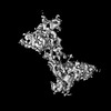 36730MC M: map data used to model this data C: citing same article ( |
|---|---|
| Similar structure data |


-Links
 PDBj
PDBj


- Assembly
| Deposited unit | 
|
|---|---|
| 1 |
|
-Components
| #1: Antibody | Mass: 15539.212 Da / Num. of mol.: 2 Source method: isolated from a genetically manipulated source Source: (gene. exp.)  Homo sapiens (human) Homo sapiens (human)#2: Protein | Mass: 22169.053 Da / Num. of mol.: 2 Source method: isolated from a genetically manipulated source Source: (gene. exp.) Severe acute respiratory syndrome coronavirus 2Gene: S, 2 / Production host: Homo sapiens (human) / References: UniProt: P0DTC2Has protein modification | Y | |
|---|
-Experimental details
-Experiment
| Experiment | Method: ELECTRON MICROSCOPY |
|---|---|
| EM experiment | Aggregation state: PARTICLE / 3D reconstruction method: single particle reconstruction |
- Sample preparation
| Component | Name: complex of RBD domain of SARS-CoV-2 spike protein and IBT-CoV144 Type: COMPLEX / Entity ID: all / Source: MULTIPLE SOURCES |
|---|---|
| Source (natural) | Organism: |
| Source (recombinant) | Organism: Homo sapiens (human) |
| Buffer solution | pH: 7.4 |
| Specimen | Conc.: 1.2 mg/ml / Embedding applied: NO / Shadowing applied: NO / Staining applied: NO / Vitrification applied: YES |
| Specimen support | Grid material: COPPER / Grid mesh size: 300 divisions/in. / Grid type: Quantifoil R1.2/1.3 |
| Vitrification | Instrument: FEI VITROBOT MARK IV / Cryogen name: ETHANE / Humidity: 100 % / Chamber temperature: 277 K |
- Electron microscopy imaging
| Experimental equipment |  Model: Titan Krios / Image courtesy: FEI Company |
|---|---|
| Microscopy | Model: FEI TITAN KRIOS |
| Electron gun | Electron source:  FIELD EMISSION GUN / Accelerating voltage: 300 kV / Illumination mode: FLOOD BEAM FIELD EMISSION GUN / Accelerating voltage: 300 kV / Illumination mode: FLOOD BEAM |
| Electron lens | Mode: BRIGHT FIELD / Nominal defocus max: 2500 nm / Nominal defocus min: 1500 nm / Calibrated defocus min: 1500 nm / Calibrated defocus max: 2500 nm / Cs: 2.7 mm / C2 aperture diameter: 70 µm / Alignment procedure: COMA FREE |
| Specimen holder | Cryogen: NITROGEN / Specimen holder model: FEI TITAN KRIOS AUTOGRID HOLDER / Temperature (max): 93 K / Temperature (min): 93 K |
| Image recording | Electron dose: 50 e/Å2 / Detector mode: COUNTING / Film or detector model: GATAN K2 SUMMIT (4k x 4k) |
- Processing
| EM software |
| ||||||||||||||||||||||||||||||||
|---|---|---|---|---|---|---|---|---|---|---|---|---|---|---|---|---|---|---|---|---|---|---|---|---|---|---|---|---|---|---|---|---|---|
| CTF correction | Type: PHASE FLIPPING AND AMPLITUDE CORRECTION | ||||||||||||||||||||||||||||||||
| 3D reconstruction | Resolution: 4.5 Å / Resolution method: FSC 0.143 CUT-OFF / Num. of particles: 203158 / Num. of class averages: 1 / Symmetry type: POINT | ||||||||||||||||||||||||||||||||
| Refine LS restraints |
|
