 Movie
Movie Controller
 Structure viewers
 About EMN search


-Search query
-Search result
Showing 1 - 50 of 215 items for (author: hansen & g)
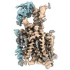
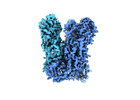
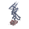
EMDB-17377: 
Structure of human SIT1 (focussed map / refinement)

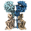
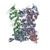
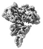
EMDB-17378:
Structure of human SIT1:ACE2 complex (open PD conformation)

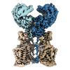
EMDB-17379:
Structure of human SIT1:ACE2 complex (closed PD conformation)
EMDB-17380:
Structure of human SIT1 bound to L-pipecolate (focussed map / refinement)
EMDB-17381:
Structure of human SIT1:ACE2 complex (open PD conformation) bound to L-pipecolate
EMDB-17382:
Structure of human SIT1:ACE2 complex (closed PD conformation) bound to L-pipecolate

PDB-8p2w:
Structure of human SIT1 (focussed map / refinement)

PDB-8p2x:
Structure of human SIT1:ACE2 complex (open PD conformation)

PDB-8p2y:
Structure of human SIT1:ACE2 complex (closed PD conformation)

PDB-8p2z:
Structure of human SIT1 bound to L-pipecolate (focussed map / refinement)
PDB-8p30:
Structure of human SIT1:ACE2 complex (open PD conformation) bound to L-pipecolate

PDB-8p31:
Structure of human SIT1:ACE2 complex (closed PD conformation) bound to L-pipecolate

EMDB-43753:
Yeast U1 snRNP with humanized U1C Zinc-Finger domain

PDB-8w2o:
Yeast U1 snRNP with humanized U1C Zinc-Finger domain
EMDB-41363:
Cryo-EM structure of DDB1deltaB-DDA1-DCAF5

PDB-8tl6:
Cryo-EM structure of DDB1deltaB-DDA1-DCAF5

EMDB-18207:
Ubiquitin ligation to substrate by a cullin-RING E3 ligase & Cdc34: NEDD8-CUL2-RBX1-ELOB/C-FEM1C with trapped UBE2R2~donor UB-Sil1 peptide, Glacios map

EMDB-18230:
Ubiquitin ligation to substrate by a cullin-RING E3 ligase & Cdc34: NEDD8-CUL2-RBX1-ELOB/C-FEM1C with trapped UBE2R2~donor UB-Sil1 peptide
EMDB-18915:
Ubiquitin ligation to neosubstrate by a cullin-RING E3 ligase & Cdc34: NEDD8-CUL2-RBX1-ELOB/C-VHL-MZ1 with trapped UBE2R2~donor UB-BRD4 BD2

PDB-8q7r:
Ubiquitin ligation to substrate by a cullin-RING E3 ligase & Cdc34: NEDD8-CUL2-RBX1-ELOB/C-FEM1C with trapped UBE2R2~donor UB-Sil1 peptide

PDB-8r5h:
Ubiquitin ligation to neosubstrate by a cullin-RING E3 ligase & Cdc34: NEDD8-CUL2-RBX1-ELOB/C-VHL-MZ1 with trapped UBE2R2~donor UB-BRD4 BD2

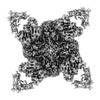
EMDB-17412:
Vaccinia Virus flower-shaped pore-like structure
EMDB-40984:
5TU-t1 - heterodimeric triplet polymerase ribozyme

PDB-8t2p:
5TU-t1 - heterodimeric triplet polymerase ribozyme
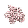
EMDB-17410:
Vaccinia Virus palisade layer A10 trimer

EMDB-17411:
Subtomogram averaging structure of the Vaccinia Virus core palisade layer

EMDB-17413:
Cryo-electron tomogram of intact mature Vaccinia Virus particle

EMDB-17414:
Cryo-electron tomogram of isolated Vaccinia Virus cores

EMDB-18452:
Vaccinia virus inner core wall
PDB-8p4k:
Vaccinia Virus palisade layer A10 trimer

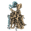
EMDB-16024:
SARS-CoV-2 S protein in complex with pT1644 Fab

EMDB-16026:
SARS-CoV-2 S protein in complex with pT1696 Fab
PDB-8bg6:
SARS-CoV-2 S protein in complex with pT1644 Fab
PDB-8bg8:
SARS-CoV-2 S protein in complex with pT1696 Fab

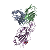
EMDB-16416:
HB3VAR03 apo headstructure (PfEMP1 A) complexed with EPCR
PDB-8c44:
HB3VAR03 apo headstructure (PfEMP1 A) complexed with EPCR
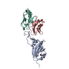
EMDB-27920:
3H03 Fab in complex with influenza virus neuraminidase from A/Brevig Mission/1/1918 (H1N1)

EMDB-27921:
2H08 Fab in complex with influenza virus neuraminidase from A/Brevig Mission/1/1918 (H1N1)

PDB-8e6j:
3H03 Fab in complex with influenza virus neuraminidase from A/Brevig Mission/1/1918 (H1N1)

PDB-8e6k:
2H08 Fab in complex with influenza virus neuraminidase from A/Brevig Mission/1/1918 (H1N1)
EMDB-16415:
HB3VAR03 apo headstructure (PfEMP1 A)

PDB-8c3y:
HB3VAR03 apo headstructure (PfEMP1 A)

EMDB-27738:
Negative stain EM map of the heterodimeric p110gamma-p84 complex
EMDB-27627:
Structure of p110 gamma bound to the Ras inhibitory nanobody NB7
PDB-8dp0:
Structure of p110 gamma bound to the Ras inhibitory nanobody NB7

EMDB-26989:
Extracellular architecture of an engineered canonical Wnt signaling ternary complex

PDB-8ctg:
Extracellular architecture of an engineered canonical Wnt signaling ternary complex

EMDB-25699:
VFLIP Spike Trimer with GAR03

EMDB-25700:
VFLIP Spike Trimer with GAR05 FAB

PDB-7t5o:
VFLIP Spike Trimer with GAR03
Pages:
 wwPDB to switch to version 3 of the EMDB data model
