 Movie
Movie Controller

[English] 日本語
 Yorodumi
- EMDB-27627: Structure of p110 gamma bound to the Ras inhibitory nanobody NB7 -
+ Open data

- Basic information
| Entry |  | |||||||||
|---|---|---|---|---|---|---|---|---|---|---|
| Title | Structure of p110 gamma bound to the Ras inhibitory nanobody NB7 | |||||||||
 Map data Map data | human p110 gamma bound to Nanobody NB8 | |||||||||
 Sample Sample |
| |||||||||
 Keywords Keywords | PI3K / p110 / PIK3CG / phosphoinositide 3-kinase / PIP3 / TRANSFERASE-IMMUNE SYSTEM complex | |||||||||
| Function / homology |  Function and homology information Function and homology informationnegative regulation of fibroblast apoptotic process / secretory granule localization / negative regulation of cardiac muscle contraction / natural killer cell chemotaxis / respiratory burst involved in defense response / neutrophil extravasation / phosphatidylinositol-4-phosphate 3-kinase / negative regulation of triglyceride catabolic process / positive regulation of acute inflammatory response / T cell chemotaxis ...negative regulation of fibroblast apoptotic process / secretory granule localization / negative regulation of cardiac muscle contraction / natural killer cell chemotaxis / respiratory burst involved in defense response / neutrophil extravasation / phosphatidylinositol-4-phosphate 3-kinase / negative regulation of triglyceride catabolic process / positive regulation of acute inflammatory response / T cell chemotaxis / regulation of calcium ion transmembrane transport / phosphatidylinositol 3-kinase complex, class IB / regulation of cell adhesion mediated by integrin / Co-stimulation by ICOS / T cell proliferation / sphingosine-1-phosphate receptor signaling pathway / 1-phosphatidylinositol-4-phosphate 3-kinase activity / phosphatidylinositol 3-kinase complex, class IA / phosphatidylinositol-4,5-bisphosphate 3-kinase / 1-phosphatidylinositol-4,5-bisphosphate 3-kinase activity / phosphatidylinositol 3-kinase / phosphatidylinositol-3-phosphate biosynthetic process / hepatocyte apoptotic process / dendritic cell chemotaxis / 1-phosphatidylinositol-3-kinase activity / Erythropoietin activates Phosphoinositide-3-kinase (PI3K) / positive regulation of MAP kinase activity / phosphatidylinositol phosphate biosynthetic process / Synthesis of PIPs at the plasma membrane / phosphatidylinositol-mediated signaling / regulation of angiogenesis / CD28 dependent PI3K/Akt signaling / positive regulation of Rac protein signal transduction / ephrin receptor binding / GPVI-mediated activation cascade / neutrophil chemotaxis / positive regulation of endothelial cell migration / cellular response to cAMP / positive regulation of cytokine production / phosphatidylinositol 3-kinase/protein kinase B signal transduction / mast cell degranulation / T cell activation / platelet aggregation / endocytosis / Constitutive Signaling by Aberrant PI3K in Cancer / phospholipase C-activating G protein-coupled receptor signaling pathway / PIP3 activates AKT signaling / G beta:gamma signalling through PI3Kgamma / cell migration / positive regulation of cytosolic calcium ion concentration / PI5P, PP2A and IER3 Regulate PI3K/AKT Signaling / angiogenesis / adaptive immune response / positive regulation of phosphatidylinositol 3-kinase/protein kinase B signal transduction / protein kinase activity / non-specific serine/threonine protein kinase / immune response / inflammatory response / G protein-coupled receptor signaling pathway / innate immune response / protein serine kinase activity / protein serine/threonine kinase activity / ATP binding / membrane / identical protein binding / plasma membrane / cytosol / cytoplasm Similarity search - Function | |||||||||
| Biological species |  Homo sapiens (human) / Homo sapiens (human) /  | |||||||||
| Method | single particle reconstruction / cryo EM / Resolution: 2.96 Å | |||||||||
 Authors Authors | Burke JE / Nam SE / Rathinaswamy MK / Yip CK | |||||||||
| Funding support |  Canada, 1 items Canada, 1 items
| |||||||||
 Citation Citation | Journal: Elife / Year: 2023 Title: Allosteric activation or inhibition of PI3Kγ mediated through conformational changes in the p110γ helical domain. Authors: Noah J Harris / Meredith L Jenkins / Sung-Eun Nam / Manoj K Rathinaswamy / Matthew A H Parson / Harish Ranga-Prasad / Udit Dalwadi / Brandon E Moeller / Eleanor Sheeky / Scott D Hansen / ...Authors: Noah J Harris / Meredith L Jenkins / Sung-Eun Nam / Manoj K Rathinaswamy / Matthew A H Parson / Harish Ranga-Prasad / Udit Dalwadi / Brandon E Moeller / Eleanor Sheeky / Scott D Hansen / Calvin K Yip / John E Burke /  Abstract: PI3Kγ is a critical immune signaling enzyme activated downstream of diverse cell surface molecules, including Ras, PKCβ activated by the IgE receptor, and Gβγ subunits released from activated ...PI3Kγ is a critical immune signaling enzyme activated downstream of diverse cell surface molecules, including Ras, PKCβ activated by the IgE receptor, and Gβγ subunits released from activated GPCRs. PI3Kγ can form two distinct complexes, with the p110γ catalytic subunit binding to either a p101 or p84 regulatory subunit, with these complexes being differentially activated by upstream stimuli. Here, using a combination of cryo electron microscopy, HDX-MS, and biochemical assays, we have identified novel roles of the helical domain of p110γ in regulating lipid kinase activity of distinct PI3Kγ complexes. We defined the molecular basis for how an allosteric inhibitory nanobody potently inhibits kinase activity through rigidifying the helical domain and regulatory motif of the kinase domain. The nanobody did not block either p110γ membrane recruitment or Ras/Gβγ binding, but instead decreased ATP turnover. We also identified that p110γ can be activated by dual PKCβ helical domain phosphorylation leading to partial unfolding of an N-terminal region of the helical domain. PKCβ phosphorylation is selective for p110γ-p84 compared to p110γ-p101, driven by differential dynamics of the helical domain of these different complexes. Nanobody binding prevented PKCβ-mediated phosphorylation. Overall, this work shows an unexpected allosteric regulatory role of the helical domain of p110γ that is distinct between p110γ-p84 and p110γ-p101 and reveals how this can be modulated by either phosphorylation or allosteric inhibitory binding partners. This opens possibilities of future allosteric inhibitor development for therapeutic intervention. | |||||||||
| History |
|
- Structure visualization
| Supplemental images |
|---|

- Downloads & links
-EMDB archive
| Map data | emd_27627.map.gz | 188.7 MB | EMDB map data format | |
|---|---|---|---|---|
| Header (meta data) | emd-27627-v30.xmlemd-27627.xml | 18.3 KB 18.3 KB | Display Display | EMDB header |
| Images | 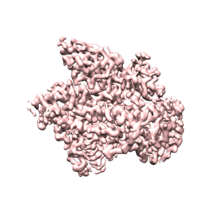 emd_27627.png emd_27627.png | 70.5 KB | ||
| Filedesc metadata | emd-27627.cif.gz | 7.4 KB | ||
| Archive directory |  http://ftp.pdbj.org/pub/emdb/structures/EMD-27627ftp://ftp.pdbj.org/pub/emdb/structures/EMD-27627 http://ftp.pdbj.org/pub/emdb/structures/EMD-27627ftp://ftp.pdbj.org/pub/emdb/structures/EMD-27627 | HTTPS FTP |
-Related structure data
| Related structure data | 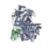 8dp0MC M: atomic model generated by this map C: citing same article ( |
|---|---|
| Similar structure data |
-Links
| EMDB pages | EMDB (EBI/PDBe) / EMDataResource |
|---|---|
| Related items in Molecule of the Month |


-Map
| File | Download / File: emd_27627.map.gz / Format: CCP4 / Size: 209.3 MB / Type: IMAGE STORED AS FLOATING POINT NUMBER (4 BYTES) | ||||||||||||||||||||||||||||||||||||
|---|---|---|---|---|---|---|---|---|---|---|---|---|---|---|---|---|---|---|---|---|---|---|---|---|---|---|---|---|---|---|---|---|---|---|---|---|---|
| Annotation | human p110 gamma bound to Nanobody NB8 | ||||||||||||||||||||||||||||||||||||
| Projections & slices | Image control
Images are generated by Spider. | ||||||||||||||||||||||||||||||||||||
| Voxel size | X=Y=Z: 0.83 Å | ||||||||||||||||||||||||||||||||||||
| Density |
| ||||||||||||||||||||||||||||||||||||
| Symmetry | Space group: 1 | ||||||||||||||||||||||||||||||||||||
| Details | EMDB XML:
|
 Z (Sec.)
 Y (Row.)
 X (Col.)


-Supplemental data
- Sample components
-Entire : Complex of p110 gamma with NB7
| Entire | Name: Complex of p110 gamma with NB7 |
|---|---|
| Components |
|
-Supramolecule #1: Complex of p110 gamma with NB7
| Supramolecule | Name: Complex of p110 gamma with NB7 / type: complex / ID: 1 / Parent: 0 / Macromolecule list: all |
|---|---|
| Source (natural) | Organism: Homo sapiens (human) |
| Molecular weight | Theoretical: 126.454 KDa |
-Macromolecule #1: Phosphatidylinositol 4,5-bisphosphate 3-kinase catalytic subunit ...
| Macromolecule | Name: Phosphatidylinositol 4,5-bisphosphate 3-kinase catalytic subunit gamma isoform type: protein_or_peptide / ID: 1 / Number of copies: 1 / Enantiomer: LEVO / EC number: phosphatidylinositol 3-kinase |
|---|---|
| Source (natural) | Organism: Homo sapiens (human) |
| Molecular weight | Theoretical: 126.627406 KDa |
| Recombinant expression | Organism:   Spodoptera frugiperda (fall armyworm) Spodoptera frugiperda (fall armyworm) |
| Sequence | String: MELENYKQPV VLREDNCRRR RRMKPRSAAA SLSSMELIPI EFVLPTSQRK CKSPETALLH VAGHGNVEQM KAQVWLRALE TSVAADFYH RLGPHHFLLL YQKKGQWYEI YDKYQVVQTL DCLRYWKATH RSPGQIHLVQ RHPPSEESQA FQRQLTALIG Y DVTDVSNV ...String: MELENYKQPV VLREDNCRRR RRMKPRSAAA SLSSMELIPI EFVLPTSQRK CKSPETALLH VAGHGNVEQM KAQVWLRALE TSVAADFYH RLGPHHFLLL YQKKGQWYEI YDKYQVVQTL DCLRYWKATH RSPGQIHLVQ RHPPSEESQA FQRQLTALIG Y DVTDVSNV HDDELEFTRR GLVTPRMAEV ASRDPKLYAM HPWVTSKPLP EYLWKKIANN CIFIVIHRST TSQTIKVSPD DT PGAILQS FFTKMAKKKS LMDIPESQSE QDFVLRVCGR DEYLVGETPI KNFQWVRHCL KNGEEIHVVL DTPPDPALDE VRK EEWPLV DDCTGVTGYH EQLTIHGKDH ESVFTVSLWD CDRKFRVKIR GIDIPVLPRN TDLTVFVEAN IQHGQQVLCQ RRTS PKPFT EEVLWNVWLE FSIKIKDLPK GALLNLQIYC GKAPALSSKA SAESPSSESK GKVQLLYYVN LLLIDHRFLL RRGEY VLHM WQISGKGEDQ GSFNADKLTS ATNPDKENSM SISILLDNYC HPIALPKHQP TPDPEGDRVR AEMPNQLRKQ LEAIIA TDP LNPLTAEDKE LLWHFRYESL KHPKAYPKLF SSVKWGQQEI VAKTYQLLAR REVWDQSALD VGLTMQLLDC NFSDENV RA IAVQKLESLE DDDVLHYLLQ LVQAVKFEPY HDSALARFLL KRGLRNKRIG HFLFWFLRSE IAQSRHYQQR FAVILEAY L RGCGTAMLHD FTQQVQVIEM LQKVTLDIKS LSAEKYDVSS QVISQLKQKL ENLQNSQLPE SFRVPYDPGL KAGALAIEK CKVMASKKKP LWLEFKCADP TALSNETIGI IFKHGDDLRQ DMLILQILRI MESIWETESL DLCLLPYGCI STGDKIGMIE IVKDATTIA KIQQSTVGNT GAFKDEVLNH WLKEKSPTEE KFQAAVERFV YSCAGYCVAT FVLGIGDRHN DNIMITETGN L FHIDFGHI LGNYKSFLGI NKERVPFVLT PDFLFVMGTS GKKTSPHFQK FQDICVKAYL ALRHHTNLLI ILFSMMLMTG MP QLTSKED IEYIRDALTV GKNEEDAKKY FLDQIEVCRD KGWTVQFNWF LHLVLGIKQG EKHSA UniProtKB: Phosphatidylinositol 4,5-bisphosphate 3-kinase catalytic subunit gamma isoform |
-Macromolecule #2: Nanobody NB7
| Macromolecule | Name: Nanobody NB7 / type: protein_or_peptide / ID: 2 / Number of copies: 1 / Enantiomer: LEVO |
|---|---|
| Source (natural) | Organism: |
| Molecular weight | Theoretical: 14.574995 KDa |
| Recombinant expression | Organism:  |
| Sequence | String: QVQLVESGGG LVQPGGSLRL SCAASGSIFS INAMGWYRQA PGKQRELVAH ITSGGSTNYA DSVKGRFTIS RDNAKNTVYL QMNSLKPED TAVYYCNEAG DPFLGSTWNG PPAFGSWGQG TQVTVSSHHH HHHEPEA |
-Experimental details
-Structure determination
| Method | cryo EM |
|---|---|
 Processing Processing | single particle reconstruction |
| Aggregation state | particle |
-Sample preparation
| Concentration | 0.45 mg/mL | |||||||||||||||
|---|---|---|---|---|---|---|---|---|---|---|---|---|---|---|---|---|
| Buffer | pH: 8.5 Component:
Details: Freshly prepared gel filtration buffer, filtered through 0.22 um filter and degassed | |||||||||||||||
| Grid | Model: C-flat-2/2 / Material: COPPER / Mesh: 300 / Pretreatment - Type: GLOW DISCHARGE / Pretreatment - Time: 25 sec. / Pretreatment - Atmosphere: AIR / Pretreatment - Pressure: 0.039 kPa | |||||||||||||||
| Vitrification | Cryogen name: ETHANE / Chamber humidity: 100 % / Chamber temperature: 277.15 K / Instrument: FEI VITROBOT MARK IV Details: 3 uL of specimen was loaded onto grid and immediately blotted for 1.5 seconds with -5 force before plunging into liquid ethane.. | |||||||||||||||
| Details | Specimen was p110 gamma purified with stabilizing nanobody, NB7, to homogeneity by gel filtration. |
- Electron microscopy
| Microscope | FEI TITAN KRIOS |
|---|---|
| Image recording | Film or detector model: GATAN K3 BIOQUANTUM (6k x 4k) / Number grids imaged: 1 / Number real images: 7344 / Average electron dose: 50.0 e/Å2 |
| Electron beam | Acceleration voltage: 300 kV / Electron source:  FIELD EMISSION GUN FIELD EMISSION GUN |
| Electron optics | Illumination mode: FLOOD BEAM / Imaging mode: BRIGHT FIELD / Nominal defocus max: 2.5 µm / Nominal defocus min: 0.5 µm |
| Sample stage | Specimen holder model: FEI TITAN KRIOS AUTOGRID HOLDER / Cooling holder cryogen: NITROGEN |
| Experimental equipment |  Model: Titan Krios / Image courtesy: FEI Company |

