 Movie
Movie Controller


+ Open data

- Basic information
| Entry | Database: PDB / ID: 8dp0 | |||||||||||||||||||||
|---|---|---|---|---|---|---|---|---|---|---|---|---|---|---|---|---|---|---|---|---|---|---|
| Title | Structure of p110 gamma bound to the Ras inhibitory nanobody NB7 | |||||||||||||||||||||
 Components Components |
| |||||||||||||||||||||
 Keywords Keywords | TRANSFERASE/IMMUNE SYSTEM / PI3K / p110 / PIK3CG / phosphoinositide 3-kinase / PIP3 / TRANSFERASE-IMMUNE SYSTEM complex | |||||||||||||||||||||
| Function / homology |  Function and homology information Function and homology informationnegative regulation of fibroblast apoptotic process / secretory granule localization / negative regulation of cardiac muscle contraction / natural killer cell chemotaxis / respiratory burst involved in defense response / neutrophil extravasation / phosphatidylinositol-4-phosphate 3-kinase / negative regulation of triglyceride catabolic process / positive regulation of acute inflammatory response / T cell chemotaxis ...negative regulation of fibroblast apoptotic process / secretory granule localization / negative regulation of cardiac muscle contraction / natural killer cell chemotaxis / respiratory burst involved in defense response / neutrophil extravasation / phosphatidylinositol-4-phosphate 3-kinase / negative regulation of triglyceride catabolic process / positive regulation of acute inflammatory response / T cell chemotaxis / regulation of calcium ion transmembrane transport / phosphatidylinositol 3-kinase complex, class IB / regulation of cell adhesion mediated by integrin / Co-stimulation by ICOS / T cell proliferation / sphingosine-1-phosphate receptor signaling pathway / 1-phosphatidylinositol-4-phosphate 3-kinase activity / phosphatidylinositol 3-kinase complex, class IA / phosphatidylinositol-4,5-bisphosphate 3-kinase / 1-phosphatidylinositol-4,5-bisphosphate 3-kinase activity / phosphatidylinositol 3-kinase / phosphatidylinositol-3-phosphate biosynthetic process / hepatocyte apoptotic process / dendritic cell chemotaxis / 1-phosphatidylinositol-3-kinase activity / Erythropoietin activates Phosphoinositide-3-kinase (PI3K) / positive regulation of MAP kinase activity / phosphatidylinositol phosphate biosynthetic process / Synthesis of PIPs at the plasma membrane / phosphatidylinositol-mediated signaling / regulation of angiogenesis / CD28 dependent PI3K/Akt signaling / positive regulation of Rac protein signal transduction / ephrin receptor binding / GPVI-mediated activation cascade / neutrophil chemotaxis / positive regulation of endothelial cell migration / cellular response to cAMP / positive regulation of cytokine production / phosphatidylinositol 3-kinase/protein kinase B signal transduction / mast cell degranulation / T cell activation / platelet aggregation / endocytosis / Constitutive Signaling by Aberrant PI3K in Cancer / phospholipase C-activating G protein-coupled receptor signaling pathway / PIP3 activates AKT signaling / G beta:gamma signalling through PI3Kgamma / cell migration / positive regulation of cytosolic calcium ion concentration / PI5P, PP2A and IER3 Regulate PI3K/AKT Signaling / angiogenesis / adaptive immune response / positive regulation of phosphatidylinositol 3-kinase/protein kinase B signal transduction / protein kinase activity / non-specific serine/threonine protein kinase / immune response / inflammatory response / G protein-coupled receptor signaling pathway / innate immune response / protein serine kinase activity / protein serine/threonine kinase activity / ATP binding / membrane / identical protein binding / plasma membrane / cytosol / cytoplasm Similarity search - Function | |||||||||||||||||||||
| Biological species |  Homo sapiens (human) Homo sapiens (human) | |||||||||||||||||||||
| Method | ELECTRON MICROSCOPY / single particle reconstruction / cryo EM / Resolution: 2.96 Å | |||||||||||||||||||||
 Authors Authors | Burke, J.E. / Nam, S.E. / Rathinaswamy, M.K. / Yip, C.K. | |||||||||||||||||||||
| Funding support |  Canada, 1items Canada, 1items
| |||||||||||||||||||||
 Citation Citation | Journal: Elife / Year: 2023 Title: Allosteric activation or inhibition of PI3Kγ mediated through conformational changes in the p110γ helical domain. Authors: Noah J Harris / Meredith L Jenkins / Sung-Eun Nam / Manoj K Rathinaswamy / Matthew A H Parson / Harish Ranga-Prasad / Udit Dalwadi / Brandon E Moeller / Eleanor Sheeky / Scott D Hansen / ...Authors: Noah J Harris / Meredith L Jenkins / Sung-Eun Nam / Manoj K Rathinaswamy / Matthew A H Parson / Harish Ranga-Prasad / Udit Dalwadi / Brandon E Moeller / Eleanor Sheeky / Scott D Hansen / Calvin K Yip / John E Burke /  Abstract: PI3Kγ is a critical immune signaling enzyme activated downstream of diverse cell surface molecules, including Ras, PKCβ activated by the IgE receptor, and Gβγ subunits released from activated ...PI3Kγ is a critical immune signaling enzyme activated downstream of diverse cell surface molecules, including Ras, PKCβ activated by the IgE receptor, and Gβγ subunits released from activated GPCRs. PI3Kγ can form two distinct complexes, with the p110γ catalytic subunit binding to either a p101 or p84 regulatory subunit, with these complexes being differentially activated by upstream stimuli. Here, using a combination of cryo electron microscopy, HDX-MS, and biochemical assays, we have identified novel roles of the helical domain of p110γ in regulating lipid kinase activity of distinct PI3Kγ complexes. We defined the molecular basis for how an allosteric inhibitory nanobody potently inhibits kinase activity through rigidifying the helical domain and regulatory motif of the kinase domain. The nanobody did not block either p110γ membrane recruitment or Ras/Gβγ binding, but instead decreased ATP turnover. We also identified that p110γ can be activated by dual PKCβ helical domain phosphorylation leading to partial unfolding of an N-terminal region of the helical domain. PKCβ phosphorylation is selective for p110γ-p84 compared to p110γ-p101, driven by differential dynamics of the helical domain of these different complexes. Nanobody binding prevented PKCβ-mediated phosphorylation. Overall, this work shows an unexpected allosteric regulatory role of the helical domain of p110γ that is distinct between p110γ-p84 and p110γ-p101 and reveals how this can be modulated by either phosphorylation or allosteric inhibitory binding partners. This opens possibilities of future allosteric inhibitor development for therapeutic intervention. | |||||||||||||||||||||
| History |
|
- Structure visualization
| Structure viewer | Molecule: MolmilJmol/JSmol |
|---|
- Downloads & links
-Download
| PDBx/mmCIF format | 8dp0.cif.gz | 208.2 KB | Display | PDBx/mmCIF format |
|---|---|---|---|---|
| PDB format | pdb8dp0.ent.gz | 162.3 KB | Display | PDB format |
| PDBx/mmJSON format | 8dp0.json.gz | Tree view | PDBx/mmJSON format | |
| Others |  Other downloads Other downloads |
-Validation report
| Arichive directory | https://data.pdbj.org/pub/pdb/validation_reports/dp/8dp0ftp://data.pdbj.org/pub/pdb/validation_reports/dp/8dp0 | HTTPS FTP |
|---|
-Related structure data
| Related structure data |  27627MC M: map data used to model this data C: citing same article ( |
|---|---|
| Similar structure data |
-Links
 PDBj
PDBj


- Assembly
| Deposited unit | 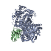
|
|---|---|
| 1 |
|
-Components
| #1: Protein | Mass: 126627.406 Da / Num. of mol.: 1 Source method: isolated from a genetically manipulated source Source: (gene. exp.) Homo sapiens (human) / Gene: PIK3CG / Production host:   Spodoptera frugiperda (fall armyworm) Spodoptera frugiperda (fall armyworm)References: UniProt: P48736, phosphatidylinositol 3-kinase, phosphatidylinositol-4,5-bisphosphate 3-kinase, phosphatidylinositol-4-phosphate 3-kinase, non-specific serine/threonine protein kinase |
|---|---|
| #2: Antibody | Mass: 14574.995 Da / Num. of mol.: 1 Source method: isolated from a genetically manipulated source Source: (gene. exp.)  |
| Has protein modification | N |
-Experimental details
-Experiment
| Experiment | Method: ELECTRON MICROSCOPY |
|---|---|
| EM experiment | Aggregation state: PARTICLE / 3D reconstruction method: single particle reconstruction |
- Sample preparation
| Component | Name: Complex of p110 gamma with NB7 / Type: COMPLEX / Entity ID: all / Source: MULTIPLE SOURCES | |||||||||||||||||||||||||
|---|---|---|---|---|---|---|---|---|---|---|---|---|---|---|---|---|---|---|---|---|---|---|---|---|---|---|
| Molecular weight | Value: 0.126454 MDa / Experimental value: NO | |||||||||||||||||||||||||
| Source (natural) | Organism: Homo sapiens (human) | |||||||||||||||||||||||||
| Source (recombinant) | Organism: Spodoptera frugiperda (fall armyworm) | |||||||||||||||||||||||||
| Buffer solution | pH: 8.5 Details: Freshly prepared gel filtration buffer, filtered through 0.22 um filter and degassed | |||||||||||||||||||||||||
| Buffer component |
| |||||||||||||||||||||||||
| Specimen | Conc.: 0.45 mg/ml / Embedding applied: NO / Shadowing applied: NO / Staining applied: NO / Vitrification applied: YES Details: Specimen was p110 gamma purified with stabilizing nanobody, NB7, to homogeneity by gel filtration. | |||||||||||||||||||||||||
| Specimen support | Grid material: COPPER / Grid mesh size: 300 divisions/in. / Grid type: C-flat-2/2 | |||||||||||||||||||||||||
| Vitrification | Instrument: FEI VITROBOT MARK IV / Cryogen name: ETHANE / Humidity: 100 % / Chamber temperature: 277.15 K Details: 3 uL of specimen was loaded onto grid and immediately blotted for 1.5 seconds with -5 force before plunging into liquid ethane. |
- Electron microscopy imaging
| Experimental equipment |  Model: Titan Krios / Image courtesy: FEI Company |
|---|---|
| Microscopy | Model: FEI TITAN KRIOS |
| Electron gun | Electron source:  FIELD EMISSION GUN / Accelerating voltage: 300 kV / Illumination mode: FLOOD BEAM FIELD EMISSION GUN / Accelerating voltage: 300 kV / Illumination mode: FLOOD BEAM |
| Electron lens | Mode: BRIGHT FIELD / Nominal defocus max: 2500 nm / Nominal defocus min: 500 nm |
| Specimen holder | Cryogen: NITROGEN / Specimen holder model: FEI TITAN KRIOS AUTOGRID HOLDER |
| Image recording | Electron dose: 50 e/Å2 / Film or detector model: GATAN K3 BIOQUANTUM (6k x 4k) / Num. of grids imaged: 1 / Num. of real images: 7344 |
- Processing
| Software | Name: PHENIX / Version: 1.19.1_4122: / Classification: refinement | ||||||||||||||||||||||||||||||||
|---|---|---|---|---|---|---|---|---|---|---|---|---|---|---|---|---|---|---|---|---|---|---|---|---|---|---|---|---|---|---|---|---|---|
| EM software |
| ||||||||||||||||||||||||||||||||
| CTF correction | Type: PHASE FLIPPING AND AMPLITUDE CORRECTION | ||||||||||||||||||||||||||||||||
| Symmetry | Point symmetry: C1 (asymmetric) | ||||||||||||||||||||||||||||||||
| 3D reconstruction | Resolution: 2.96 Å / Resolution method: FSC 0.143 CUT-OFF / Num. of particles: 149603 / Num. of class averages: 1 / Symmetry type: POINT | ||||||||||||||||||||||||||||||||
| Atomic model building | PDB-ID: 7MEZ Pdb chain-ID: A / Accession code: 7MEZ / Pdb chain residue range: 1-1102 / Source name: PDB / Type: experimental model | ||||||||||||||||||||||||||||||||
| Refine LS restraints |
|

