 ムービー
ムービー コントローラー
 構造ビューア
 EMN検索について


-検索条件
-検索結果
検索 (著者・登録者: gruene & t)の結果全42件を表示しています

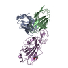
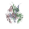
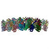
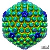
EMDB-16024: 
SARS-CoV-2 S protein in complex with pT1644 Fab

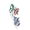
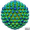
EMDB-16026:
SARS-CoV-2 S protein in complex with pT1696 Fab
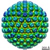
PDB-8bg6:
SARS-CoV-2 S protein in complex with pT1644 Fab
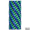
PDB-8bg8:
SARS-CoV-2 S protein in complex with pT1696 Fab
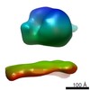
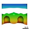
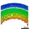
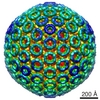
EMDB-15627:
Tomogram of the Mimivirus genomic fiber
EMDB-15628:
Tomogram of the Mimivirus genomic fiber
EMDB-15629:
Tomogram of the Mimivirus genomic fiber
EMDB-15630:
Tomogram of the Mimivirus genomic fiber

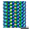
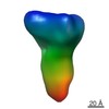
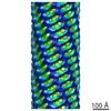
EMDB-14167:
Amyloid fibril from the antimicrobial peptide uperin 3.5

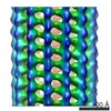
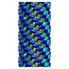
PDB-7qv5:
Amyloid fibril from the antimicrobial peptide uperin 3.5

PDB-7qv6:
Amyloid fibril from the antimicrobial peptide aurein 3.3
PDB-7bho:
DNA origami signpost designed model
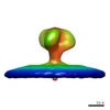
EMDB-11123:
Pre-fusion conformation of glycoprotein B of Herpes simplex virus 1
PDB-6z9m:
Pseudoatomic model of the pre-fusion conformation of glycoprotein B of Herpes simplex virus 1

PDB-6y6k:
Cryo-EM structure of a Phenuiviridae L protein

EMDB-3909:
Stalled E. coli ribosomes (Fo-c SecM nascent chains) and native E. coli membranes containing recombinant YidC

EMDB-3919:
Stalled E. coli ribosomes (Fo-c SecM nascent chains) and native E. coli membranes containing recombinant YidC

EMDB-3920:
Stalled E. coli ribosomes (Fo-c SecM nascent chains) and native E. coli membranes containing recombinant YidC

EMDB-3921:
Stalled E. coli ribosomes (Fo-c SecM nascent chains) and native E. coli membranes containing recombinant YidC

EMDB-3922:
Stalled E. coli ribosomes (Fo-c SecM nascent chains) and native E. coli membranes containing recombinant YidC

EMDB-3923:
Stalled E. coli ribosomes (Fo-c SecM nascent chains) and native E. coli membranes containing recombinant YidC
EMDB-3924:
Stalled E. coli ribosomes (Fo-c SecM nascent chains) and native E. coli membranes containing recombinant YidC

PDB-5o4w:
Protein structure determination by electron diffraction using a single three-dimensional nanocrystal

PDB-5o4x:
Protein structure determination by electron diffraction using a single three-dimensional nanocrystal
EMDB-4043:
13-protofilament microtubule structure determined in situ from U87MG cell
EMDB-4045:
13-protofilament microtubule structure determined in situ from U2OS cell
PDB-5fki:
Pseudorabies virus (PrV) nuclear egress complex proteins fitted as a hexameric lattice into a sub-tomogram average derived from focused- ion beam milled lamellae electron cryo-microscopic data
EMDB-3197:
Sub-tomogram averaging of electron cryo-microscopic data taken from focused-ion beam milled lamellae of nuclei of Pseudorabies virus (PrV) nuclear egress complex-expressing cells
EMDB-3215:
Sub-tomogram averaging of electron cryo-microscopic data taken from focused-ion beam milled lamellae of nuclei of Pseudorabies virus (PrV) nuclear egress complex-expressing cells

EMDB-2531:
Tomographic subvolume average of EFF-1 fusogen on extracellular vesicles
EMDB-2532:
Tomographic subvolume average of EFF-1 fusogen on extracellular vesicles
EMDB-1956:
Cryo Electron Tomography of Herpes Simplex Virus during Axonal Transport and Secondary Envelopment in Primary Neurons
EMDB-1957:
Cryo Electron Tomography of Herpes Simplex Virus during Axonal Transport and Secondary Envelopment in Primary Neurons
EMDB-1958:
Cryo Electron Tomography of Herpes Simplex Virus during Axonal Transport and Secondary Envelopment in Primary Neurons

EMDB-1959:
Cryo Electron Tomography of Herpes Simplex Virus during Axonal Transport and Secondary Envelopment in Primary Neurons
EMDB-1865:
Structure of the yeast eisosome core component Lsp1 filament
EMDB-1866:
Structure of the yeast eisosome core component Lsp1 filament

EMDB-1867:
Structure of the yeast eisosome core component Lsp1 filament bound to a liposome membrane.
EMDB-1868:
Structure of the yeast eisosome core component Pil1 filament bound to a liposome membrane.
EMDB-1549:
Three-dimensional icosahedral reconstruction of Rift Valley Fever virus at pH 6.0

EMDB-1550:
Three-dimensional icosahedral reconstruction of Rift Valley Fever virus at pH 7.4
EMDB-1216:
Cryo-electron tomographic structure of an immunodeficiency virus envelope complex in situ.
 wwPDBはEMDBデータモデルのバージョン3へ移行します
