 Movie
Movie Controller

[English] 日本語
 Yorodumi
- PDB-6z9m: Pseudoatomic model of the pre-fusion conformation of glycoprotein... -
+ Open data

- Basic information
| Entry | Database: PDB / ID: 6z9m | |||||||||||||||
|---|---|---|---|---|---|---|---|---|---|---|---|---|---|---|---|---|
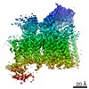
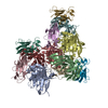
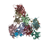
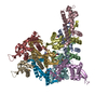
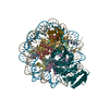
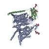
| Title | Pseudoatomic model of the pre-fusion conformation of glycoprotein B of Herpes simplex virus 1 | |||||||||||||||
 Components Components | Envelope glycoprotein B | |||||||||||||||
 Keywords Keywords | VIRAL PROTEIN / Membrane fusion protein / glycoprotein / gB / UL27 / viral entry protein / class III fusion protein / transmembrane protein / pre-fusion conformation | |||||||||||||||
| Function / homology |  Function and homology information Function and homology informationhost cell Golgi membrane / host cell endosome membrane / viral envelope / symbiont entry into host cell / virion attachment to host cell / host cell plasma membrane / virion membrane / identical protein binding Similarity search - Function | |||||||||||||||
| Biological species |   Human herpesvirus 1 (Herpes simplex virus type 1) Human herpesvirus 1 (Herpes simplex virus type 1) | |||||||||||||||
| Method | ELECTRON MICROSCOPY / subtomogram averaging / cryo EM / Resolution: 9.1 Å | |||||||||||||||
 Authors Authors | Vollmer, B. / Prazak, V. / Vasishtan, D. / Jefferys, E.E. / Hernandez-Duran, A. / Vallbracht, M. / Klupp, B. / Mettenleiter, T.C. / Backovic, M. / Rey, F.A. ...Vollmer, B. / Prazak, V. / Vasishtan, D. / Jefferys, E.E. / Hernandez-Duran, A. / Vallbracht, M. / Klupp, B. / Mettenleiter, T.C. / Backovic, M. / Rey, F.A. / Topf, M. / Gruenewald, K. | |||||||||||||||
| Funding support |  Germany, Germany,  United Kingdom, European Union, 4items United Kingdom, European Union, 4items
| |||||||||||||||
 Citation Citation | Journal: Sci Adv / Year: 2020 Title: The prefusion structure of herpes simplex virus glycoprotein B. Authors: B Vollmer / V Pražák / D Vasishtan / E E Jefferys / A Hernandez-Duran / M Vallbracht / B G Klupp / T C Mettenleiter / M Backovic / F A Rey / M Topf / K Grünewald /  Abstract: Cell entry of enveloped viruses requires specialized viral proteins that mediate fusion with the host membrane by substantial structural rearrangements from a metastable pre- to a stable postfusion ...Cell entry of enveloped viruses requires specialized viral proteins that mediate fusion with the host membrane by substantial structural rearrangements from a metastable pre- to a stable postfusion conformation. This metastability renders the herpes simplex virus 1 (HSV-1) fusion glycoprotein B (gB) highly unstable such that it readily converts into the postfusion form, thereby precluding structural elucidation of the pharmacologically relevant prefusion conformation. By identification of conserved sequence signatures and molecular dynamics simulations, we devised a mutation that stabilized this form. Functionally locking gB allowed the structural determination of its membrane-embedded prefusion conformation at sub-nanometer resolution and enabled the unambiguous fit of all ectodomains. The resulting pseudo-atomic model reveals a notable conservation of conformational domain rearrangements during fusion between HSV-1 gB and the vesicular stomatitis virus glycoprotein G, despite their very distant phylogeny. In combination with our comparative sequence-structure analysis, these findings suggest common fusogenic domain rearrangements in all class III viral fusion proteins. | |||||||||||||||
| History |
|
- Structure visualization
| Movie |
Movie viewer |
|---|---|
| Structure viewer | Molecule: MolmilJmol/JSmol |
- Downloads & links
-Download
| PDBx/mmCIF format | 6z9m.cif.gz | 344.7 KB | Display | PDBx/mmCIF format |
|---|---|---|---|---|
| PDB format | pdb6z9m.ent.gz | 273.1 KB | Display | PDB format |
| PDBx/mmJSON format | 6z9m.json.gz | Tree view | PDBx/mmJSON format | |
| Others |  Other downloads Other downloads |
-Validation report
| Arichive directory | https://data.pdbj.org/pub/pdb/validation_reports/z9/6z9mftp://data.pdbj.org/pub/pdb/validation_reports/z9/6z9m | HTTPS FTP |
|---|
-Related structure data
| Related structure data | 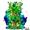 11123MC M: map data used to model this data C: citing same article ( |
|---|---|
| Similar structure data |


-Links
 PDBj
PDBj- Assembly
| Deposited unit | 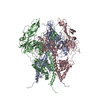
|
|---|---|
| 1 |
|
-Components
| #1: Protein | Mass: 100383.305 Da / Num. of mol.: 3 / Mutation: H516P Source method: isolated from a genetically manipulated source Source: (gene. exp.) Human herpesvirus 1 (Herpes simplex virus type 1)Gene: UL27, gB, HHV1gp041 / Details (production host): derived from pEP98 / Cell (production host): Fibroblast / Production host:  Mesocricetus auratus (golden hamster) / References: UniProt: A1Z0P7, UniProt: P06437*PLUS Mesocricetus auratus (golden hamster) / References: UniProt: A1Z0P7, UniProt: P06437*PLUSHas protein modification | Y | |
|---|
-Experimental details
-Experiment
| Experiment | Method: ELECTRON MICROSCOPY |
|---|---|
| EM experiment | Aggregation state: PARTICLE / 3D reconstruction method: subtomogram averaging |
- Sample preparation
| Component | Name: Human alphaherpesvirus 1 / Type: VIRUS Details: Protein recombinantly expressed in membrane protein enriched extracellular vesicles (MPEEVs) Entity ID: all / Source: RECOMBINANT | |||||||||||||||
|---|---|---|---|---|---|---|---|---|---|---|---|---|---|---|---|---|
| Molecular weight | Value: 0.3 MDa / Experimental value: NO | |||||||||||||||
| Source (natural) | Organism: Human alphaherpesvirus 1 (Herpes simplex virus type 1) | |||||||||||||||
| Source (recombinant) | Organism: Mesocricetus auratus (golden hamster) / Cell: Fibroblast | |||||||||||||||
| Details of virus | Empty: YES / Enveloped: YES / Isolate: OTHER / Type: VIRUS-LIKE PARTICLE | |||||||||||||||
| Natural host | Organism: Homo sapiens | |||||||||||||||
| Buffer solution | pH: 7.8 | |||||||||||||||
| Buffer component |
| |||||||||||||||
| Specimen | Embedding applied: NO / Shadowing applied: NO / Staining applied: NO / Vitrification applied: YES Details: Protein recombinantly expressed in membrane protein enriched extracellular vesicles (MPEEVs) | |||||||||||||||
| Specimen support | Grid material: COPPER / Grid type: Quantifoil R2/1 | |||||||||||||||
| Vitrification | Instrument: HOMEMADE PLUNGER / Cryogen name: ETHANE-PROPANE |
- Electron microscopy imaging
| Experimental equipment |  Model: Tecnai Polara / Image courtesy: FEI Company |
|---|---|
| Microscopy | Model: FEI POLARA 300 Details: Additional Dataset collected using Titan Krios (FEI Thermo) at 300 kV with a 70 um C2 aperture and post-column QUANTUM energy filter operated in Zero-Loss mode using 20 eV energy slit and K2 ...Details: Additional Dataset collected using Titan Krios (FEI Thermo) at 300 kV with a 70 um C2 aperture and post-column QUANTUM energy filter operated in Zero-Loss mode using 20 eV energy slit and K2 Summit direct electron detector in counting mode (Gatan). Defocus range: 2300 - 4900 nm. |
| Electron gun | Electron source:  FIELD EMISSION GUN / Accelerating voltage: 300 kV / Illumination mode: FLOOD BEAM FIELD EMISSION GUN / Accelerating voltage: 300 kV / Illumination mode: FLOOD BEAM |
| Electron lens | Mode: BRIGHT FIELD / Calibrated defocus min: 1600 nm / Calibrated defocus max: 3500 nm / Cs: 2 mm / C2 aperture diameter: 70 µm |
| Specimen holder | Cryogen: NITROGEN / Specimen holder model: OTHER |
| Image recording | Electron dose: 2.3 e/Å2 / Detector mode: COUNTING / Film or detector model: GATAN K2 SUMMIT (4k x 4k) |
| EM imaging optics | Energyfilter name: GIF Quantum LS / Energyfilter slit width: 20 eV |
| Image scans | Width: 3836 / Height: 3710 |
- Processing
| EM software |
| |||||||||||||||||||||||||||
|---|---|---|---|---|---|---|---|---|---|---|---|---|---|---|---|---|---|---|---|---|---|---|---|---|---|---|---|---|
| CTF correction | Type: PHASE FLIPPING ONLY | |||||||||||||||||||||||||||
| Symmetry | Point symmetry: C3 (3 fold cyclic) | |||||||||||||||||||||||||||
| 3D reconstruction | Resolution: 9.1 Å / Resolution method: FSC 0.143 CUT-OFF / Num. of particles: 46067 / Algorithm: BACK PROJECTION / Num. of class averages: 1 / Symmetry type: POINT | |||||||||||||||||||||||||||
| EM volume selection | Method: Manual / Num. of tomograms: 99 / Num. of volumes extracted: 56176 | |||||||||||||||||||||||||||
| Atomic model building | B value: 300 / Protocol: FLEXIBLE FIT / Space: REAL / Target criteria: Cross correlation coefficient | |||||||||||||||||||||||||||
| Atomic model building | PDB-ID: 5V2S Pdb chain-ID: A / Accession code: 5V2S / Pdb chain residue range: 104-723 / Source name: PDB / Type: experimental model |

