 Movie
Movie Controller

[English] 日本語
 Yorodumi
- PDB-5v2s: Crystal structure of glycoprotein B from Herpes Simplex Virus type I -
+ Open data

- Basic information
| Entry | Database: PDB / ID: 5v2s | ||||||||||||
|---|---|---|---|---|---|---|---|---|---|---|---|---|---|
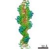
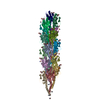
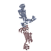
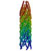
| Title | Crystal structure of glycoprotein B from Herpes Simplex Virus type I | ||||||||||||
 Components Components | Envelope glycoprotein B | ||||||||||||
 Keywords Keywords | VIRAL PROTEIN / fusogen / bitopic membrane protein | ||||||||||||
| Function / homology |  Function and homology information Function and homology informationhost cell Golgi membrane / host cell endosome membrane / viral envelope / symbiont entry into host cell / virion attachment to host cell / host cell plasma membrane / virion membrane Similarity search - Function | ||||||||||||
| Biological species |   Human herpesvirus 1 (Herpes simplex virus type 1) Human herpesvirus 1 (Herpes simplex virus type 1) | ||||||||||||
| Method |  X-RAY DIFFRACTION / SYNCHROTRON / MOLECULAR REPLACEMENT / Resolution: 3.6 Å X-RAY DIFFRACTION / SYNCHROTRON / MOLECULAR REPLACEMENT / Resolution: 3.6 Å | ||||||||||||
 Authors Authors | Cooper, R.S. / Heldwein, E.E. | ||||||||||||
| Funding support |  United States, 3items United States, 3items
| ||||||||||||
 Citation Citation | Journal: Nat. Struct. Mol. Biol. / Year: 2018 Title: Structural basis for membrane anchoring and fusion regulation of the herpes simplex virus fusogen gB. Authors: Cooper, R.S. / Georgieva, E.R. / Borbat, P.P. / Freed, J.H. / Heldwein, E.E. | ||||||||||||
| History |
|
- Structure visualization
| Structure viewer | Molecule: MolmilJmol/JSmol |
|---|
- Downloads & links
-Download
| PDBx/mmCIF format | 5v2s.cif.gz | 162 KB | Display | PDBx/mmCIF format |
|---|---|---|---|---|
| PDB format | pdb5v2s.ent.gz | 122.8 KB | Display | PDB format |
| PDBx/mmJSON format | 5v2s.json.gz | Tree view | PDBx/mmJSON format | |
| Others |  Other downloads Other downloads |
-Validation report
| Arichive directory | https://data.pdbj.org/pub/pdb/validation_reports/v2/5v2sftp://data.pdbj.org/pub/pdb/validation_reports/v2/5v2s | HTTPS FTP |
|---|
-Related structure data
| Related structure data |  6bm8C 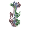 2gumS C: citing same article ( S: Starting model for refinement |
|---|---|
| Similar structure data |
-Links
 PDBj
PDBj- Assembly
| Deposited unit | 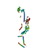
| ||||||||
|---|---|---|---|---|---|---|---|---|---|
| 1 | 
| ||||||||
| 2 |
| ||||||||
| Unit cell |
| ||||||||
| Details | Trimer as determined by gel filtration |
-Components
| #1: Protein | Mass: 94501.469 Da / Num. of mol.: 1 / Fragment: residues 72-904 Source method: isolated from a genetically manipulated source Source: (gene. exp.) Human herpesvirus 1 (Herpes simplex virus type 1)Gene: UL27, HHV1gp041 / Plasmid: pFastBac / Cell line (production host): SF9 / Production host:   Spodoptera frugiperda (fall armyworm) / References: UniProt: A1Z0P7, UniProt: P10211*PLUS Spodoptera frugiperda (fall armyworm) / References: UniProt: A1Z0P7, UniProt: P10211*PLUS | ||||
|---|---|---|---|---|---|
| #2: Polysaccharide | Source method: isolated from a genetically manipulated source #3: Sugar | ChemComp-NAG / |   Type: D-saccharide, beta linking / Mass: 221.208 Da / Num. of mol.: 1 Type: D-saccharide, beta linking / Mass: 221.208 Da / Num. of mol.: 1Source method: isolated from a genetically manipulated source Formula: C8H15NO6 Has protein modification | Y | |
-Experimental details
-Experiment
| Experiment | Method: X-RAY DIFFRACTION / Number of used crystals: 1 |
|---|
- Sample preparation
| Crystal | Density Matthews: 4.66 Å3/Da / Density % sol: 73.62 % |
|---|---|
| Crystal grow | Temperature: 295 K / Method: vapor diffusion, hanging drop Details: 16% PEG 3350, 0.1 M Na Hepes pH 7.2, 0.15 M Na formate |
-Data collection
| Diffraction | Mean temperature: 100 K |
|---|---|
| Diffraction source | Source: SYNCHROTRON / Site: APS / Beamline: 24-ID-E / Wavelength: 0.97918 Å |
| Detector | Type: ADSC QUANTUM 315 / Detector: CCD / Date: Aug 22, 2016 |
| Radiation | Protocol: SINGLE WAVELENGTH / Monochromatic (M) / Laue (L): M / Scattering type: x-ray |
| Radiation wavelength | Wavelength: 0.97918 Å / Relative weight: 1 |
| Reflection | Resolution: 3.6→108.232 Å / Num. obs: 21107 / % possible obs: 99.7 % / Redundancy: 8.4 % / CC1/2: 0.98 / Rmerge(I) obs: 0.3297 / Rpim(I) all: 0.1196 / Net I/σ(I): 6.5 |
| Reflection shell | Resolution: 3.6→3.729 Å / Redundancy: 8.5 % / Rmerge(I) obs: 1.833 / Mean I/σ(I) obs: 1.15 / Num. unique obs: 2070 / CC1/2: 0.61 / Rpim(I) all: 0.6654 / % possible all: 99.76 |
- Processing
| Software |
| ||||||||||||||||||||||||
|---|---|---|---|---|---|---|---|---|---|---|---|---|---|---|---|---|---|---|---|---|---|---|---|---|---|
| Refinement | Method to determine structure: MOLECULAR REPLACEMENT Starting model: 2GUM Resolution: 3.6→108.232 Å / SU ML: 0.57 / Cross valid method: FREE R-VALUE / σ(F): 1.35 / Phase error: 29.08 / Stereochemistry target values: ML
| ||||||||||||||||||||||||
| Solvent computation | Shrinkage radii: 0.9 Å / VDW probe radii: 1.11 Å / Solvent model: FLAT BULK SOLVENT MODEL | ||||||||||||||||||||||||
| Refinement step | Cycle: LAST / Resolution: 3.6→108.232 Å
| ||||||||||||||||||||||||
| Refine LS restraints |
|
