 Movie
Movie Controller


+ Open data

- Basic information
| Entry | Database: EMDB / ID: EMD-4196 | |||||||||
|---|---|---|---|---|---|---|---|---|---|---|
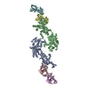
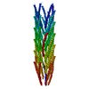
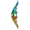
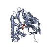
| Title | AlfA from B. subtilis plasmid pLS32 filament structure at 3.4 A | |||||||||
 Map data Map data | ||||||||||
 Sample Sample |
| |||||||||
 Keywords Keywords | plasmid segregation / bacterial cytoskeleton / cytomotive filament / VIRAL PROTEIN | |||||||||
| Function / homology | Actin-like protein, N-terminal / Actin like proteins N terminal domain / ATPase, nucleotide binding domain / Actin-like protein N-terminal domain-containing protein Function and homology information Function and homology information | |||||||||
| Biological species |  | |||||||||
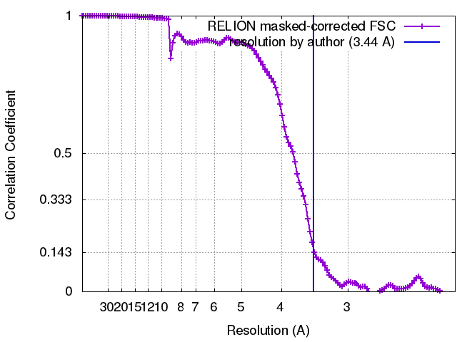
| Method | helical reconstruction / cryo EM / Resolution: 3.44 Å | |||||||||
 Authors Authors | Szewczak-Harris A / Lowe J | |||||||||
| Funding support |  United Kingdom, 1 items United Kingdom, 1 items
| |||||||||
 Citation Citation | Journal: Proc Natl Acad Sci U S A / Year: 2018 Title: Cryo-EM reconstruction of AlfA from reveals the structure of a simplified actin-like filament at 3.4-Å resolution. Authors: Andrzej Szewczak-Harris / Jan Löwe / Abstract: Low copy-number plasmid pLS32 of subsp. contains a partitioning system that ensures segregation of plasmid copies during cell division. The partitioning locus comprises actin-like protein AlfA, ...Low copy-number plasmid pLS32 of subsp. contains a partitioning system that ensures segregation of plasmid copies during cell division. The partitioning locus comprises actin-like protein AlfA, adaptor protein AlfB, and the centromeric sequence Similar to the ParMRC partitioning system from plasmid R1, AlfA filaments form actin-like double helical filaments that arrange into an antiparallel bipolar spindle, which attaches its growing ends to sister plasmids through interactions with AlfB and Because, compared with ParM and other actin-like proteins, AlfA is highly diverged in sequence, we determined the atomic structure of nonbundling AlfA filaments to 3.4-Å resolution by cryo-EM. The structure reveals how the deletion of subdomain IIB of the canonical actin fold has been accommodated by unique longitudinal and lateral contacts, while still enabling formation of left-handed, double helical, polar and staggered filaments that are architecturally similar to ParM. Through cryo-EM reconstruction of bundling AlfA filaments, we obtained a pseudoatomic model of AlfA doublets: the assembly of two filaments. The filaments are antiparallel, as required by the segregation mechanism, and exactly antiphasic with near eightfold helical symmetry, to enable efficient doublet formation. The structure of AlfA filaments and doublets shows, in atomic detail, how deletion of an entire domain of the actin fold is compensated by changes to all interfaces so that the required properties of polymerization, nucleotide hydrolysis, and antiparallel doublet formation are retained to fulfill the system's biological raison d'être. | |||||||||
| History |
|
- Structure visualization
| Movie |
Movie viewer |
|---|---|
| Structure viewer | EM map: SurfViewMolmilJmol/JSmol |
| Supplemental images |

- Downloads & links
-EMDB archive
| Map data | emd_4196.map.gz | 152.9 MB | EMDB map data format | |
|---|---|---|---|---|
| Header (meta data) | emd-4196-v30.xmlemd-4196.xml | 14.6 KB 14.6 KB | Display Display | EMDB header |
| FSC (resolution estimation) | emd_4196_fsc.xml | 12.4 KB | Display | FSC data file |
| Images | 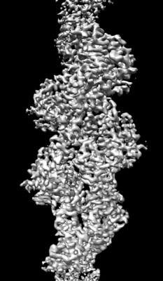 emd_4196.png emd_4196.png | 45.9 KB | ||
| Filedesc metadata | emd-4196.cif.gz | 6 KB | ||
| Archive directory |  http://ftp.pdbj.org/pub/emdb/structures/EMD-4196ftp://ftp.pdbj.org/pub/emdb/structures/EMD-4196 http://ftp.pdbj.org/pub/emdb/structures/EMD-4196ftp://ftp.pdbj.org/pub/emdb/structures/EMD-4196 | HTTPS FTP |
-Related structure data
| Related structure data | 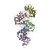 6f95MC M: atomic model generated by this map C: citing same article ( |
|---|---|
| Similar structure data |

-Links
| EMDB pages | EMDB (EBI/PDBe) / EMDataResource |
|---|
-Map
| File | Download / File: emd_4196.map.gz / Format: CCP4 / Size: 163.6 MB / Type: IMAGE STORED AS FLOATING POINT NUMBER (4 BYTES) | ||||||||||||||||||||||||||||||||||||||||||||||||||||||||||||
|---|---|---|---|---|---|---|---|---|---|---|---|---|---|---|---|---|---|---|---|---|---|---|---|---|---|---|---|---|---|---|---|---|---|---|---|---|---|---|---|---|---|---|---|---|---|---|---|---|---|---|---|---|---|---|---|---|---|---|---|---|---|
| Projections & slices | Image control
Images are generated by Spider. | ||||||||||||||||||||||||||||||||||||||||||||||||||||||||||||
| Voxel size | X=Y=Z: 1.07 Å | ||||||||||||||||||||||||||||||||||||||||||||||||||||||||||||
| Density |
| ||||||||||||||||||||||||||||||||||||||||||||||||||||||||||||
| Symmetry | Space group: 1 | ||||||||||||||||||||||||||||||||||||||||||||||||||||||||||||
| Details | EMDB XML:
CCP4 map header:
| ||||||||||||||||||||||||||||||||||||||||||||||||||||||||||||
 Z (Sec.)
 Y (Row.)
 X (Col.)


-Supplemental data
- Sample components
-Entire : AlfA filament
| Entire | Name: AlfA filament |
|---|---|
| Components |
|
-Supramolecule #1: AlfA filament
| Supramolecule | Name: AlfA filament / type: complex / ID: 1 / Parent: 0 / Macromolecule list: #1 |
|---|---|
| Source (natural) | Organism: |
-Macromolecule #1: AlfA
| Macromolecule | Name: AlfA / type: protein_or_peptide / ID: 1 / Number of copies: 5 / Enantiomer: LEVO |
|---|---|
| Source (natural) | Organism: |
| Molecular weight | Theoretical: 31.200281 KDa |
| Recombinant expression | Organism: |
| Sequence | String: GSHMTLTTVI DIGNFSTKYA YKDAAQIKVG SFPSILHSYK PLEDYEGMER VEYNGLDYYV GETVKNFYFG REEQMYFGNT RKGHMEGQI RLVYALYTIF KETGAAEFNL ILTCPYESMV TDKKYFVQHF EGEREVIVEG KSFKFTVHNI VMAAEGLGAL N FSDSLNCV ...String: GSHMTLTTVI DIGNFSTKYA YKDAAQIKVG SFPSILHSYK PLEDYEGMER VEYNGLDYYV GETVKNFYFG REEQMYFGNT RKGHMEGQI RLVYALYTIF KETGAAEFNL ILTCPYESMV TDKKYFVQHF EGEREVIVEG KSFKFTVHNI VMAAEGLGAL N FSDSLNCV IVDAGSKTLN VLYLINGSIS KMDSHTINGG TIDNSIMDLA KTFAKTCSNI DYDYPIVCTG GKAEEMKECL EN VGYSTVS SAELGEDKPS YYVNSVGLLL KYGRKFEEMF A UniProtKB: Actin-like protein N-terminal domain-containing protein |
-Macromolecule #2: MAGNESIUM ION
| Macromolecule | Name: MAGNESIUM ION / type: ligand / ID: 2 / Number of copies: 5 / Formula: MG |
|---|---|
| Molecular weight | Theoretical: 24.305 Da |
-Macromolecule #3: ADENOSINE-5'-DIPHOSPHATE
| Macromolecule | Name: ADENOSINE-5'-DIPHOSPHATE / type: ligand / ID: 3 / Number of copies: 5 / Formula: ADP |
|---|---|
| Molecular weight | Theoretical: 427.201 Da |
| Chemical component information |  ChemComp-ADP: |
-Experimental details
-Structure determination
| Method | cryo EM |
|---|---|
 Processing Processing | helical reconstruction |
| Aggregation state | filament |
-Sample preparation
| Buffer | pH: 8 Details: 20 mM Tris-HCl, 100 mM KCl, 2 mM TCEP, 1mM NaN3, pH 8.0, 5 mM ATP and 10 mM MgCl2 added to |
|---|---|
| Grid | Model: Quantifoil R1.2/1.3 / Material: COPPER / Mesh: 300 |
| Vitrification | Cryogen name: ETHANE / Instrument: FEI VITROBOT MARK IV |
- Electron microscopy
| Microscope | FEI TITAN KRIOS |
|---|---|
| Image recording | Film or detector model: FEI FALCON III (4k x 4k) / Detector mode: COUNTING / Number real images: 368 / Average electron dose: 33.0 e/Å2 |
| Electron beam | Acceleration voltage: 300 kV / Electron source:  FIELD EMISSION GUN FIELD EMISSION GUN |
| Electron optics | Illumination mode: FLOOD BEAM / Imaging mode: BRIGHT FIELD |
| Experimental equipment |  Model: Titan Krios / Image courtesy: FEI Company |
+Image processing
-Atomic model buiding 1
| Refinement | Protocol: AB INITIO MODEL |
|---|---|
| Output model | PDB-6f95: |
