 Movie
Movie Controller


+ Open data

- Basic information
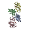
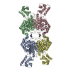
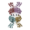
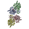
| Entry | Database: PDB / ID: 6f95 | ||||||||||||||||||||||||||||||||||||||||||
|---|---|---|---|---|---|---|---|---|---|---|---|---|---|---|---|---|---|---|---|---|---|---|---|---|---|---|---|---|---|---|---|---|---|---|---|---|---|---|---|---|---|---|---|
| Title | AlfA from B. subtilis plasmid pLS32 filament structure at 3.4 A | ||||||||||||||||||||||||||||||||||||||||||
 Components Components | AlfA | ||||||||||||||||||||||||||||||||||||||||||
 Keywords Keywords | VIRAL PROTEIN / plasmid segregation / bacterial cytoskeleton / cytomotive filament | ||||||||||||||||||||||||||||||||||||||||||
| Function / homology | Actin-like protein, N-terminal / Actin like proteins N terminal domain / ATPase, nucleotide binding domain / ATPase, nucleotide binding domain / Nucleotidyltransferase; domain 5 / 2-Layer Sandwich / Alpha Beta / ADENOSINE-5'-DIPHOSPHATE / Actin-like protein N-terminal domain-containing protein Function and homology information Function and homology information | ||||||||||||||||||||||||||||||||||||||||||
| Biological species |  | ||||||||||||||||||||||||||||||||||||||||||
| Method | ELECTRON MICROSCOPY / helical reconstruction / cryo EM / Resolution: 3.44 Å | ||||||||||||||||||||||||||||||||||||||||||
 Authors Authors | Szewczak-Harris, A. / Lowe, J. | ||||||||||||||||||||||||||||||||||||||||||
| Funding support |  United Kingdom, 1items United Kingdom, 1items
| ||||||||||||||||||||||||||||||||||||||||||
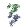
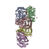
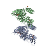
 Citation Citation | Journal: Proc Natl Acad Sci U S A / Year: 2018 Title: Cryo-EM reconstruction of AlfA from reveals the structure of a simplified actin-like filament at 3.4-Å resolution. Authors: Andrzej Szewczak-Harris / Jan Löwe / Abstract: Low copy-number plasmid pLS32 of subsp. contains a partitioning system that ensures segregation of plasmid copies during cell division. The partitioning locus comprises actin-like protein AlfA, ...Low copy-number plasmid pLS32 of subsp. contains a partitioning system that ensures segregation of plasmid copies during cell division. The partitioning locus comprises actin-like protein AlfA, adaptor protein AlfB, and the centromeric sequence Similar to the ParMRC partitioning system from plasmid R1, AlfA filaments form actin-like double helical filaments that arrange into an antiparallel bipolar spindle, which attaches its growing ends to sister plasmids through interactions with AlfB and Because, compared with ParM and other actin-like proteins, AlfA is highly diverged in sequence, we determined the atomic structure of nonbundling AlfA filaments to 3.4-Å resolution by cryo-EM. The structure reveals how the deletion of subdomain IIB of the canonical actin fold has been accommodated by unique longitudinal and lateral contacts, while still enabling formation of left-handed, double helical, polar and staggered filaments that are architecturally similar to ParM. Through cryo-EM reconstruction of bundling AlfA filaments, we obtained a pseudoatomic model of AlfA doublets: the assembly of two filaments. The filaments are antiparallel, as required by the segregation mechanism, and exactly antiphasic with near eightfold helical symmetry, to enable efficient doublet formation. The structure of AlfA filaments and doublets shows, in atomic detail, how deletion of an entire domain of the actin fold is compensated by changes to all interfaces so that the required properties of polymerization, nucleotide hydrolysis, and antiparallel doublet formation are retained to fulfill the system's biological raison d'être. | ||||||||||||||||||||||||||||||||||||||||||
| History |
|
- Structure visualization
| Movie |
Movie viewer |
|---|---|
| Structure viewer | Molecule: MolmilJmol/JSmol |
- Downloads & links
-Download
| PDBx/mmCIF format | 6f95.cif.gz | 237.7 KB | Display | PDBx/mmCIF format |
|---|---|---|---|---|
| PDB format | pdb6f95.ent.gz | 193.3 KB | Display | PDB format |
| PDBx/mmJSON format | 6f95.json.gz | Tree view | PDBx/mmJSON format | |
| Others |  Other downloads Other downloads |
-Validation report
| Arichive directory | https://data.pdbj.org/pub/pdb/validation_reports/f9/6f95ftp://data.pdbj.org/pub/pdb/validation_reports/f9/6f95 | HTTPS FTP |
|---|
-Related structure data
| Related structure data | 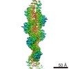 4196MC M: map data used to model this data C: citing same article ( |
|---|---|
| Similar structure data |

-Links
 PDBj
PDBj- Assembly
| Deposited unit | 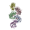
|
|---|---|
| 1 |
|
-Components
| #1: Protein | Mass: 31200.281 Da / Num. of mol.: 5 / Mutation: K21A, K22A, K101A, K102A Source method: isolated from a genetically manipulated source Source: (gene. exp.) #2: Chemical | ChemComp-MG /   Mass: 24.305 Da / Num. of mol.: 5 / Source method: obtained synthetically / Formula: Mg Mass: 24.305 Da / Num. of mol.: 5 / Source method: obtained synthetically / Formula: Mg#3: Chemical | ChemComp-ADP /   Mass: 427.201 Da / Num. of mol.: 5 / Source method: obtained synthetically / Formula: C10H15N5O10P2 / Comment: ADP, energy-carrying molecule*YM Mass: 427.201 Da / Num. of mol.: 5 / Source method: obtained synthetically / Formula: C10H15N5O10P2 / Comment: ADP, energy-carrying molecule*YMHas protein modification | N | |
|---|
-Experimental details
-Experiment
| Experiment | Method: ELECTRON MICROSCOPY |
|---|---|
| EM experiment | Aggregation state: FILAMENT / 3D reconstruction method: helical reconstruction |
- Sample preparation
| Component | Name: AlfA filament / Type: COMPLEX / Entity ID: #1 / Source: RECOMBINANT |
|---|---|
| Source (natural) | Organism: |
| Source (recombinant) | Organism: |
| Buffer solution | pH: 8 Details: 20 mM Tris-HCl, 100 mM KCl, 2 mM TCEP, 1mM NaN3, pH 8.0, 5 mM ATP and 10 mM MgCl2 added to |
| Specimen | Embedding applied: NO / Shadowing applied: NO / Staining applied: NO / Vitrification applied: YES |
| Specimen support | Grid material: COPPER / Grid mesh size: 300 divisions/in. / Grid type: Quantifoil R1.2/1.3 |
| Vitrification | Instrument: FEI VITROBOT MARK IV / Cryogen name: ETHANE |
- Electron microscopy imaging
| Experimental equipment |  Model: Titan Krios / Image courtesy: FEI Company |
|---|---|
| Microscopy | Model: FEI TITAN KRIOS |
| Electron gun | Electron source:  FIELD EMISSION GUN / Accelerating voltage: 300 kV / Illumination mode: FLOOD BEAM FIELD EMISSION GUN / Accelerating voltage: 300 kV / Illumination mode: FLOOD BEAM |
| Electron lens | Mode: BRIGHT FIELD |
| Image recording | Electron dose: 33 e/Å2 / Detector mode: COUNTING / Film or detector model: FEI FALCON III (4k x 4k) / Num. of real images: 368 |
- Processing
| Software | Name: PHENIX / Version: dev_2919: / Classification: refinement | ||||||||||||||||||||||||
|---|---|---|---|---|---|---|---|---|---|---|---|---|---|---|---|---|---|---|---|---|---|---|---|---|---|
| EM software | Name: PHENIX / Category: model refinement | ||||||||||||||||||||||||
| CTF correction | Type: PHASE FLIPPING AND AMPLITUDE CORRECTION | ||||||||||||||||||||||||
| Helical symmerty | Angular rotation/subunit: 156.521 ° / Axial rise/subunit: 24.9219 Å / Axial symmetry: C1 | ||||||||||||||||||||||||
| 3D reconstruction | Resolution: 3.44 Å / Resolution method: FSC 0.143 CUT-OFF / Num. of particles: 78787 / Symmetry type: HELICAL | ||||||||||||||||||||||||
| Atomic model building | Protocol: AB INITIO MODEL | ||||||||||||||||||||||||
| Refine LS restraints |
|
