 Movie
Movie Controller

[English] 日本語
 Yorodumi
- PDB-2hil: Structure of the Neisseria gonorrhoeae Type IV pilus filament fro... -
+ Open data

- Basic information
| Entry | Database: PDB / ID: 2hil | |||||||||
|---|---|---|---|---|---|---|---|---|---|---|
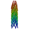
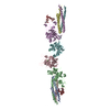
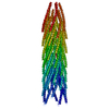
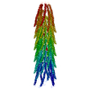
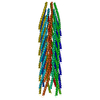
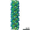
| Title | Structure of the Neisseria gonorrhoeae Type IV pilus filament from x-ray crystallography and electron cryomicroscopy | |||||||||
 Components Components | Fimbrial protein | |||||||||
 Keywords Keywords | CELL ADHESION / TYPE IV PILI / virulence factors / DNA binding protein / natural transformation / antigenic variation | |||||||||
| Function / homology |  Function and homology information Function and homology information | |||||||||
| Biological species |  Neisseria gonorrhoeae (bacteria) Neisseria gonorrhoeae (bacteria) | |||||||||
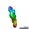
| Method | ELECTRON MICROSCOPY / helical reconstruction / cryo EM / Resolution: 12.5 Å | |||||||||
 Authors Authors | Craig, L. / Volkmann, N. / Egelman, E.H. / Tainer, J.A. | |||||||||
 Citation Citation | Journal: Mol Cell / Year: 2006 Title: Type IV pilus structure by cryo-electron microscopy and crystallography: implications for pilus assembly and functions. Authors: Lisa Craig / Niels Volkmann / Andrew S Arvai / Michael E Pique / Mark Yeager / Edward H Egelman / John A Tainer /  Abstract: Type IV pili (T4P) are long, thin, flexible filaments on bacteria that undergo assembly-disassembly from inner membrane pilin subunits and exhibit astonishing multifunctionality. Neisseria ...Type IV pili (T4P) are long, thin, flexible filaments on bacteria that undergo assembly-disassembly from inner membrane pilin subunits and exhibit astonishing multifunctionality. Neisseria gonorrhoeae (gonococcal or GC) T4P are prototypic virulence factors and immune targets for increasingly antibiotic-resistant human pathogens, yet detailed structures are unavailable for any T4P. Here, we determined a detailed experimental GC-T4P structure by quantitative fitting of a 2.3 A full-length pilin crystal structure into a 12.5 A resolution native GC-T4P reconstruction solved by cryo-electron microscopy (cryo-EM) and iterative helical real space reconstruction. Spiraling three-helix bundles form the filament core, anchor the globular heads, and provide strength and flexibility. Protruding hypervariable loops and posttranslational modifications in the globular head shield conserved functional residues in pronounced grooves, creating a surprisingly corrugated pilus surface. These results clarify T4P multifunctionality and assembly-disassembly while suggesting unified assembly mechanisms for T4P, archaeal flagella, and type II secretion system filaments. | |||||||||
| History |
|
- Structure visualization
| Movie |
Movie viewer |
|---|---|
| Structure viewer | Molecule: MolmilJmol/JSmol |
- Downloads & links
-Download
| PDBx/mmCIF format | 2hil.cif.gz | 541.5 KB | Display | PDBx/mmCIF format |
|---|---|---|---|---|
| PDB format | pdb2hil.ent.gz | 446.4 KB | Display | PDB format |
| PDBx/mmJSON format | 2hil.json.gz | Tree view | PDBx/mmJSON format | |
| Others |  Other downloads Other downloads |
-Validation report
| Arichive directory | https://data.pdbj.org/pub/pdb/validation_reports/hi/2hilftp://data.pdbj.org/pub/pdb/validation_reports/hi/2hil | HTTPS FTP |
|---|
-Related structure data
| Related structure data | 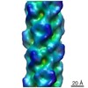 1236MC 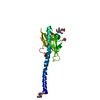 2hi2C C: citing same article ( M: map data used to model this data |
|---|---|
| Similar structure data |


-Links
 PDBj
PDBj
- Assembly
| Deposited unit | 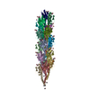
|
|---|---|
| 1 |
|
-Components
| #1: Protein | Mass: 17210.494 Da / Num. of mol.: 18 / Source method: isolated from a natural source / Source: (natural) Neisseria gonorrhoeae (bacteria) / References: UniProt: P02974#2: Polysaccharide | alpha-D-galactopyranose-(1-3)-2,4-bisacetamido-2,4-dideoxy-beta-D-glucopyranose Source method: isolated from a genetically manipulated source #3: Chemical | ChemComp-OPE / 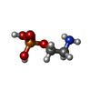  Mass: 141.063 Da / Num. of mol.: 18 / Source method: obtained synthetically / Formula: C2H8NO4P Mass: 141.063 Da / Num. of mol.: 18 / Source method: obtained synthetically / Formula: C2H8NO4PHas protein modification | Y | Sequence details | ACCORDING TO AUTHORS THE RESIDUES HAVE BEEN CONFIRMED BY SEQUENCING THE PILE GENE THAT ENCODES THE ...ACCORDING TO AUTHORS THE RESIDUES HAVE BEEN CONFIRMED BY SEQUENCING | |
|---|
-Experimental details
-Experiment
| Experiment | Method: ELECTRON MICROSCOPY |
|---|---|
| EM experiment | Aggregation state: FILAMENT / 3D reconstruction method: helical reconstruction |
- Sample preparation
| Component | Name: N. gonorrhoeae Type IV pili / Type: COMPLEX Details: native pili were sheared from N. gonorrhoeae cells and purified by successive rounds of precipitation in neutral pH buffer, which causes the filaments to aggregate, followed by resuspension in high pH buffer |
|---|---|
| Buffer solution | Name: 50 mM CHES / pH: 9.5 / Details: 50 mM CHES |
| Specimen | Conc.: 0.1 mg/ml / Embedding applied: NO / Shadowing applied: NO / Staining applied: NO / Vitrification applied: YES |
| Specimen support | Details: Quantifoil holey carbon grids, glow discharged with amyl amine |
| Vitrification | Instrument: FEI VITROBOT MARK I / Cryogen name: ETHANE Details: 5 ul of sample were applied to grids for 1 minute, blotted for 2.5 seconds then plunge-frozen in liquid ethane using an FEI Vitrobot |
- Electron microscopy imaging
| Microscopy | Model: FEI/PHILIPS CM200FEG / Date: Jun 23, 2004 |
|---|---|
| Electron gun | Electron source:  FIELD EMISSION GUN / Accelerating voltage: 120 kV / Illumination mode: FLOOD BEAM FIELD EMISSION GUN / Accelerating voltage: 120 kV / Illumination mode: FLOOD BEAM |
| Electron lens | Mode: BRIGHT FIELD / Nominal magnification: 50000 X / Nominal defocus max: 2500 nm / Nominal defocus min: 1100 nm |
| Specimen holder | Temperature: 95 K / Tilt angle max: 0 ° / Tilt angle min: 0 ° |
| Image recording | Electron dose: 10 e/Å2 / Film or detector model: KODAK SO-163 FILM |
| Image scans | Num. digital images: 12 |
| Radiation | Protocol: SINGLE WAVELENGTH / Monochromatic (M) / Laue (L): M |
| Radiation wavelength | Relative weight: 1 |
- Processing
| EM software |
| ||||||||||||
|---|---|---|---|---|---|---|---|---|---|---|---|---|---|
| CTF correction | Details: Wiener filter | ||||||||||||
| 3D reconstruction | Method: IHRSR / Resolution: 12.5 Å / Num. of particles: 25000 / Nominal pixel size: 2.23676682 Å / Actual pixel size: 2.23676682 Å / Symmetry type: HELICAL | ||||||||||||
| Atomic model building | Protocol: RIGID BODY FIT / Space: REAL / Target criteria: real-space density correlation coefficient / Details: METHOD--CoAn REFINEMENT PROTOCOL--rigid body | ||||||||||||
| Atomic model building | PDB-ID: 2HI2 Accession code: 2HI2 / Source name: PDB / Type: experimental model | ||||||||||||
| Refinement | Highest resolution: 12.5 Å Details: The GC pilus filament structure was built by computationally fitting a single GC pilin subunit (2HI2.pdb) into a cryoEM density map of the pilus filament (EMDB code xxx) using the program ...Details: The GC pilus filament structure was built by computationally fitting a single GC pilin subunit (2HI2.pdb) into a cryoEM density map of the pilus filament (EMDB code xxx) using the program CoAn (Volkmann and Hanein, J. Struct. Biol., 125, 176-184, 1999), and then applying the symmetry operators defined by the cryoEM reconstruction (10.5 Angstrom rise and a 100.8 degree azimuthal rotation, Craig et al., Mol. Cell xxx, yyy, 2006) to the subunit. | ||||||||||||
| Refinement step | Cycle: LAST / Highest resolution: 12.5 Å
| ||||||||||||
| NMR software | Name: CoAn / Classification: refinement | ||||||||||||
| Refinement | Software ordinal: 1 Details: The GC pilus filament structure was built by computationally fitting a single GC pilin subunit (2HI2.pdb) into a cryoEM density map of the pilus filament (EMDB code xxx) using the program ...Details: The GC pilus filament structure was built by computationally fitting a single GC pilin subunit (2HI2.pdb) into a cryoEM density map of the pilus filament (EMDB code xxx) using the program CoAn (Volkmann and Hanein, J. Struct. Biol., 125, 176-184, 1999), and then applying the symmetry operators defined by the cryoEM reconstruction (10.5 Angstrom rise and a 100.8 degree azimuthal rotation, Craig et al., Mol. Cell xxx, yyy, 2006) to the subunit. |
