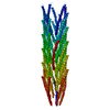
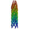
 Movie
Movie Controller

[English] 日本語
 Yorodumi
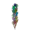
- PDB-2c0w: Molecular Structure of fd Filamentous Bacteriophage Refined with ... -
+ Open data

- Basic information
| Entry | Database: PDB / ID: 2c0w | ||||||
|---|---|---|---|---|---|---|---|
| Title | Molecular Structure of fd Filamentous Bacteriophage Refined with Respect to X-ray Fibre Diffraction | ||||||
 Components Components | COAT PROTEIN B | ||||||
 Keywords Keywords | VIRUS / CAPSID PROTEIN / FILAMENTOUS BACTERIOPHAGE / MEMBRANE PROTEIN / STRUCTURAL PROTEIN / HELICAL VIRUS | ||||||
| Function / homology |  Function and homology information Function and homology information | ||||||
| Biological species |  ENTEROBACTERIA PHAGE FD (virus) ENTEROBACTERIA PHAGE FD (virus) | ||||||
| Method |  FIBER DIFFRACTION / SYNCHROTRON / MOLECULAR REPLACEMENT / Resolution: 3.2 Å FIBER DIFFRACTION / SYNCHROTRON / MOLECULAR REPLACEMENT / Resolution: 3.2 Å | ||||||
 Authors Authors | Marvin, D.A. / Welsh, L.C. / Symmons, M.F. / Scott, W.R.P. / Straus, S.K. | ||||||
 Citation Citation | Journal: J.Mol.Biol. / Year: 2006 Title: Molecular Structure of Fd (F1, M13) Filamentous Bacteriophage Refined with Respect to X-Ray Fibre Diffraction and Solid-State NMR Data Supports Specific Models of Phage Assembly at the Bacterial Membrane. Authors: Marvin, D.A. / Welsh, L.C. / Symmons, M.F. / Scott, W.R.P. / Straus, S.K. #1: Journal: J.Mol.Biol. / Year: 1994Title: Molecular Models and Structural Comparisons of Native and Mutant Class I Filamentous Bacteriophages Ff (Fd, F1, M13), If1 and Ike Authors: Marvin, D.A. / Hale, R.D. / Nave, C. / Helmer-Citterich, M. #2: Journal: Int.J.Biol.Macromol. / Year: 1990Title: Model-Building Studies of Inovirus: Genetic Variations on a Geometric Theme Authors: Marvin, D.A. #3: Journal: J.Mol.Biol. / Year: 1995 Title: Matching Electrostatic Charge between DNA and Coat Protein in Filamentous Bacteriophage. Fibre Diffraction of Charge-Deletion Mutants. Authors: Symmons, M.F. / Welsh, L.C. / Nave, C. / Marvin, D.A. / Perham, R.N. | ||||||
| History |
|
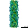
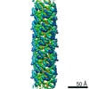
- Structure visualization
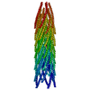
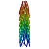
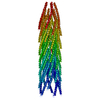
| Structure viewer | Molecule: MolmilJmol/JSmol |
|---|
- Downloads & links
-Download
| PDBx/mmCIF format | 2c0w.cif.gz | 18.7 KB | Display | PDBx/mmCIF format |
|---|---|---|---|---|
| PDB format | pdb2c0w.ent.gz | 11 KB | Display | PDB format |
| PDBx/mmJSON format | 2c0w.json.gz | Tree view | PDBx/mmJSON format | |
| Others |  Other downloads Other downloads |
-Validation report
| Arichive directory | https://data.pdbj.org/pub/pdb/validation_reports/c0/2c0wftp://data.pdbj.org/pub/pdb/validation_reports/c0/2c0w | HTTPS FTP |
|---|
-Related structure data
| Related structure data |  2c0xC  1ifjS C: citing same article ( S: Starting model for refinement |
|---|---|
| Similar structure data |
-Links
 PDBj
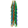
PDBj- Assembly
| Deposited unit | 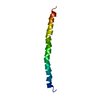
| ||||||||
|---|---|---|---|---|---|---|---|---|---|
| 1 | x 55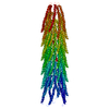
| ||||||||
| 2 |
| ||||||||
| 3 | 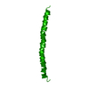
| ||||||||
| Unit cell |
| ||||||||
| Symmetry | Helical symmetry: (Circular symmetry: 5 / Dyad axis: no / N subunits divisor: 1 / Num. of operations: 55 / Rise per n subunits: 16.15 Å / Rotation per n subunits: -36 °) |
-Components
| #1: Protein/peptide | Mass: 5212.021 Da / Num. of mol.: 1 / Mutation: YES Source method: isolated from a genetically manipulated source Source: (gene. exp.) ENTEROBACTERIA PHAGE FD (virus)Description: H. HOFFMANN-BERLING Z. NATURFORSCH. SECT. B BIOSCI. V18 P876, Y1963 Production host:   PSEUDOMONAS AERUGINOSA (bacteria) / References: UniProt: P69539 PSEUDOMONAS AERUGINOSA (bacteria) / References: UniProt: P69539 |
|---|---|
| Compound details | RESIDUE IN CHAIN A, TYR 44 TO MET WAS AN INTENTIONAL MUTATION IN THE VIRAL GENOME, THIS MUTATION IS ...RESIDUE IN CHAIN A, TYR 44 TO MET WAS AN INTENTIONA |
| Sequence details | Y21M MUTANT IN CHAIN |
-Experimental details
-Experiment
| Experiment | Method: FIBER DIFFRACTION |
|---|
- Sample preparation
| Crystal grow | pH: 8 / Details: pH 8.00 |
|---|
-Data collection
| Diffraction | Mean temperature: 300 K |
|---|---|
| Diffraction source | Source: SYNCHROTRON / Site: SRS  / Beamline: PX7.2 / Wavelength: 1.488 / Beamline: PX7.2 / Wavelength: 1.488 |
| Detector | Type: MARRESEARCH / Detector: IMAGE PLATE / Details: MIRRORS |
| Radiation | Monochromator: GE CRYSTAL / Protocol: SINGLE WAVELENGTH / Monochromatic (M) / Laue (L): M / Scattering type: x-ray |
| Radiation wavelength | Wavelength: 1.488 Å / Relative weight: 1 |
| Reflection | Resolution: 3.2→44 Å / Num. obs: 0 / % possible obs: 94 % / Observed criterion σ(I): 0 |
- Processing
| Software |
| ||||||||||||||||||||||||||||||||||||||||||||||||||||||||||||
|---|---|---|---|---|---|---|---|---|---|---|---|---|---|---|---|---|---|---|---|---|---|---|---|---|---|---|---|---|---|---|---|---|---|---|---|---|---|---|---|---|---|---|---|---|---|---|---|---|---|---|---|---|---|---|---|---|---|---|---|---|---|
| Refinement | Method to determine structure: MOLECULAR REPLACEMENT Starting model: PDB ENTRY 1IFJ Resolution: 3.2→20 Å / σ(F): 0
| ||||||||||||||||||||||||||||||||||||||||||||||||||||||||||||
| Displacement parameters | Biso mean: 20 Å2 | ||||||||||||||||||||||||||||||||||||||||||||||||||||||||||||
| Refinement step | Cycle: LAST / Resolution: 3.2→20 Å
| ||||||||||||||||||||||||||||||||||||||||||||||||||||||||||||
| Refine LS restraints |
| ||||||||||||||||||||||||||||||||||||||||||||||||||||||||||||
| Xplor file | Serial no: 1 / Param file: PARALLHDG.PRO VERSION 4.05 / Topol file: TOPALLHDG.PRO VERSION 4.01 |
