 Movie
Movie Controller
 Structure viewers
 About Yorodumi Papers


+Search query
-Structure paper
| Title | Eisosome proteins assemble into a membrane scaffold. |
|---|---|
| Journal, issue, pages | J Cell Biol, Vol. 195, Issue 5, Page 889-902, Year 2011 |
| Publish date | Nov 28, 2011 |
 Authors Authors | Lena Karotki / Juha T Huiskonen / Christopher J Stefan / Natasza E Ziółkowska / Robyn Roth / Michal A Surma / Nevan J Krogan / Scott D Emr / John Heuser / Kay Grünewald / Tobias C Walther /  |
| PubMed Abstract | Spatial organization of membranes into domains of distinct protein and lipid composition is a fundamental feature of biological systems. The plasma membrane is organized in such domains to ...Spatial organization of membranes into domains of distinct protein and lipid composition is a fundamental feature of biological systems. The plasma membrane is organized in such domains to efficiently orchestrate the many reactions occurring there simultaneously. Despite the almost universal presence of membrane domains, mechanisms of their formation are often unclear. Yeast cells feature prominent plasma membrane domain organization, which is at least partially mediated by eisosomes. Eisosomes are large protein complexes that are primarily composed of many subunits of two Bin-Amphiphysin-Rvs domain-containing proteins, Pil1 and Lsp1. In this paper, we show that these proteins self-assemble into higher-order structures and bind preferentially to phosphoinositide-containing membranes. Using a combination of electron microscopy approaches, we generate structural models of Pil1 and Lsp1 assemblies, which resemble eisosomes in cells. Our data suggest that the mechanism of membrane organization by eisosomes is mediated by self-assembly of its core components into a membrane-bound protein scaffold with lipid-binding specificity. |
 External links External links | J Cell Biol / PubMed:22123866 / PubMed Central |
| Methods | EM (helical sym.) |
| Resolution | 25.0 - 31.0 Å |
| Structure data | 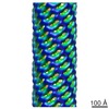 EMDB-1865: 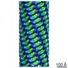 EMDB-1866:  EMDB-1867: 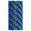 EMDB-1868: |
| Source |
|

