 ムービー
ムービー コントローラー
 構造ビューア
 EMN検索について


-検索条件
-検索結果
検索 (著者・登録者: gillet & l)の結果全46件を表示しています
EMDB-50229: 
Cryo-tomogram of FIB-milled vegetatively growing yeast cell with mitochondria
EMDB-50230:
Cryo-tomogram of FIB-milled pre-meiotic yeast cell with mitochondria
EMDB-50231:
Cryo-tomogram of FIB-milled meiotic yeast cell containing mitochondria with filaments
EMDB-50232:
Cryo-tomogram of FIB-milled yeast spore with mitochondria
EMDB-50233:
Cryo-tomogram of FIB-milled meiotic yeast cell containing mitochondria with filament arrays
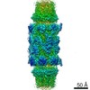
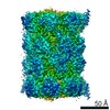
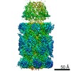
EMDB-50234:
Cryo-tomogram of purified meiotic yeast mitochondria with Ald4 filaments
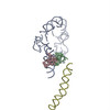
EMDB-15116:
Structural insights into the binding of bS1 to the ribosome

PDB-8a3l:
Structural insights into the binding of bS1 to the ribosome
EMDB-16556:
21S ribosomal precursors induced by heat shock.
EMDB-14571:
Negative stain 3D structure of the plastid-encoded RNA polymerase from Sinapis alba

EMDB-12261:
Structure of the HigB1 toxin mutant K95A from Mycobacterium tuberculosis (Rv1955) and its target, the cspA mRNA, on the E. coli Ribosome.

PDB-7nbu:
Structure of the HigB1 toxin mutant K95A from Mycobacterium tuberculosis (Rv1955) and its target, the cspA mRNA, on the E. coli Ribosome.
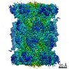
EMDB-23574:
Structure of Plasmodium falciparum 20S proteasome with bound bortezomib
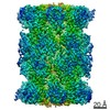
EMDB-23575:
Structure of Plasmodium falciparum 20S proteasome with bound MPI-5
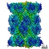
EMDB-23576:
Structure of human 20S proteasome with bound MPI-5
PDB-7lxt:
Structure of Plasmodium falciparum 20S proteasome with bound bortezomib

PDB-7lxu:
Structure of Plasmodium falciparum 20S proteasome with bound MPI-5
PDB-7lxv:
Structure of human 20S proteasome with bound MPI-5

EMDB-11710:
Structure of pre-accomodated trans-translation complex on E. coli stalled ribosome.

EMDB-11713:
Structure of accomodated trans-translation complex on E. Coli stalled ribosome.

EMDB-11717:
Structure of translocated trans-translation complex on E. coli stalled ribosome.

EMDB-11718:
Structure of post-translocated trans-translation complex on E. coli stalled ribosome.

PDB-7abz:
Structure of pre-accomodated trans-translation complex on E. coli stalled ribosome.
PDB-7ac7:
Structure of accomodated trans-translation complex on E. Coli stalled ribosome.

PDB-7acj:
Structure of translocated trans-translation complex on E. coli stalled ribosome.

PDB-7acr:
Structure of post-translocated trans-translation complex on E. coli stalled ribosome.

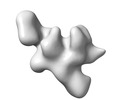
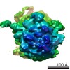
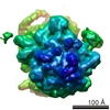
EMDB-20667:
Biochemical and structural analysis of the Neurofibromin (NF1) protein reveals high-affinity dimer formation
EMDB-20073:
Reconstruction of the Plasmodium falciparum 20S proteasome in complex with one PA28 activator prepared without chemical stabilisation
EMDB-9257:
The structure of the Plasmodium falciparum 20S proteasome in complex with two PA28 activators.
EMDB-9258:
The structure of the Plasmodium falciparum 20S proteasome.
EMDB-9259:
The structure of the Plasmodium falciparum 20S proteasome in complex with one PA28 activator.
PDB-6muv:
The structure of the Plasmodium falciparum 20S proteasome in complex with two PA28 activators
PDB-6muw:
The structure of the Plasmodium falciparum 20S proteasome.
PDB-6mux:
The structure of the Plasmodium falciparum 20S proteasome in complex with one PA28 activator
EMDB-3852:
Elongation factor G-ribosome complex captures in the absence of inhibitors.
PDB-5ot7:
Elongation factor G-ribosome complex captures in the absence of inhibitors.

PDB-3iyq:
tmRNA-SmpB: a journey to the center of the bacterial ribosome

PDB-3iyr:
tmRNA-SmpB: a journey to the center of the bacterial ribosome
EMDB-5188:
tmRNA-SmpB: a journey to the center of the bacterial ribosome
EMDB-5189:
tmRNA-SmpB: a journey to the center of the bacterial ribosome
EMDB-1312:
Scaffolding as an organizing principle in trans-translation. The roles of small protein B and ribosomal protein S1.
EMDB-1310:
Scaffolding as an organizing principle in trans-translation. The roles of small protein B and ribosomal protein S1.
PDB-2ob7:
Structure of tmRNA-(SmpB)2 complex as inferred from cryo-EM
EMDB-1311:
Cryo-EM visualization of transfer messenger RNA with two SmpBs in a stalled ribosome.
PDB-1zc8:
Coordinates of tmRNA, SmpB, EF-Tu and h44 fitted into Cryo-EM map of the 70S ribosome and tmRNA complex
EMDB-1122:
Visualizing tmRNA entry into a stalled ribosome.
 wwPDBはEMDBデータモデルのバージョン3へ移行します
