 Movie
Movie Controller

[English] 日本語
 Yorodumi
- EMDB-20073: Reconstruction of the Plasmodium falciparum 20S proteasome in com... -
+ Open data

- Basic information
| Entry | Database: EMDB / ID: EMD-20073 | |||||||||
|---|---|---|---|---|---|---|---|---|---|---|
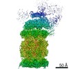
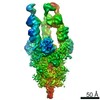
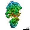
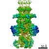
| Title | Reconstruction of the Plasmodium falciparum 20S proteasome in complex with one PA28 activator prepared without chemical stabilisation | |||||||||
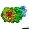
 Map data Map data | Unsharpened map | |||||||||
 Sample Sample |
| |||||||||
| Function / homology |  Function and homology information Function and homology informationER-Phagosome pathway / Cross-presentation of soluble exogenous antigens (endosomes) / Proteasome assembly / Antigen processing: Ub, ATP-independent proteasomal degradation / proteasome activator complex / Orc1 removal from chromatin / CDK-mediated phosphorylation and removal of Cdc6 / FBXL7 down-regulates AURKA during mitotic entry and in early mitosis / KEAP1-NFE2L2 pathway / UCH proteinases ...ER-Phagosome pathway / Cross-presentation of soluble exogenous antigens (endosomes) / Proteasome assembly / Antigen processing: Ub, ATP-independent proteasomal degradation / proteasome activator complex / Orc1 removal from chromatin / CDK-mediated phosphorylation and removal of Cdc6 / FBXL7 down-regulates AURKA during mitotic entry and in early mitosis / KEAP1-NFE2L2 pathway / UCH proteinases / Ub-specific processing proteases / Neddylation / Antigen processing: Ubiquitination & Proteasome degradation / MAPK6/MAPK4 signaling / AUF1 (hnRNP D0) binds and destabilizes mRNA / ABC-family protein mediated transport / Neutrophil degranulation / proteasome core complex / regulation of G1/S transition of mitotic cell cycle / proteasomal ubiquitin-independent protein catabolic process / proteasome endopeptidase complex / proteasome core complex, beta-subunit complex / endopeptidase activator activity / threonine-type endopeptidase activity / proteasome core complex, alpha-subunit complex / regulation of proteasomal protein catabolic process / proteasomal protein catabolic process / endopeptidase activity / ubiquitin-dependent protein catabolic process / proteasome-mediated ubiquitin-dependent protein catabolic process / hydrolase activity / nucleoplasm / nucleus / cytoplasm / cytosol Similarity search - Function | |||||||||
| Biological species |  | |||||||||
| Method | single particle reconstruction / cryo EM / Resolution: 4.6 Å | |||||||||
 Authors Authors | Hanssen E / Xie SC / Leis A / Metcalfe RD / Gillett DL / Tilley L / Griffin MDW | |||||||||
| Funding support |  Australia, 2 items Australia, 2 items
| |||||||||
 Citation Citation | Journal: Nat Microbiol / Year: 2019 Title: The structure of the PA28-20S proteasome complex from Plasmodium falciparum and implications for proteostasis. Authors: Stanley C Xie / Riley D Metcalfe / Eric Hanssen / Tuo Yang / David L Gillett / Andrew P Leis / Craig J Morton / Michael J Kuiper / Michael W Parker / Natalie J Spillman / Wilson Wong / ...Authors: Stanley C Xie / Riley D Metcalfe / Eric Hanssen / Tuo Yang / David L Gillett / Andrew P Leis / Craig J Morton / Michael J Kuiper / Michael W Parker / Natalie J Spillman / Wilson Wong / Christopher Tsu / Lawrence R Dick / Michael D W Griffin / Leann Tilley /  Abstract: The activity of the proteasome 20S catalytic core is regulated by protein complexes that bind to one or both ends. The PA28 regulator stimulates 20S proteasome peptidase activity in vitro, but its ...The activity of the proteasome 20S catalytic core is regulated by protein complexes that bind to one or both ends. The PA28 regulator stimulates 20S proteasome peptidase activity in vitro, but its role in vivo remains unclear. Here, we show that genetic deletion of the PA28 regulator from Plasmodium falciparum (Pf) renders malaria parasites more sensitive to the antimalarial drug dihydroartemisinin, indicating that PA28 may play a role in protection against proteotoxic stress. The crystal structure of PfPA28 reveals a bell-shaped molecule with an inner pore that has a strong segregation of charges. Small-angle X-ray scattering shows that disordered loops, which are not resolved in the crystal structure, extend from the PfPA28 heptamer and surround the pore. Using single particle cryo-electron microscopy, we solved the structure of Pf20S in complex with one and two regulatory PfPA28 caps at resolutions of 3.9 and 3.8 Å, respectively. PfPA28 binds Pf20S asymmetrically, strongly engaging subunits on only one side of the core. PfPA28 undergoes rigid body motions relative to Pf20S. Molecular dynamics simulations support conformational flexibility and a leaky interface. We propose lateral transfer of short peptides through the dynamic interface as a mechanism facilitating the release of proteasome degradation products. | |||||||||
| History |
|
- Structure visualization
| Movie |
Movie viewer |
|---|---|
| Structure viewer | EM map: SurfViewMolmilJmol/JSmol |
| Supplemental images |

- Downloads & links
-EMDB archive
| Map data | emd_20073.map.gz | 123 MB | EMDB map data format | |
|---|---|---|---|---|
| Header (meta data) | emd-20073-v30.xmlemd-20073.xml | 24 KB 24 KB | Display Display | EMDB header |
| FSC (resolution estimation) | emd_20073_fsc.xml | 12.3 KB | Display | FSC data file |
| Images | 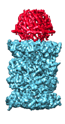 emd_20073.png emd_20073.png | 121.2 KB | ||
| Others | emd_20073_half_map_1.map.gzemd_20073_half_map_2.map.gz | 122.7 MB 122.9 MB | ||
| Archive directory |  http://ftp.pdbj.org/pub/emdb/structures/EMD-20073ftp://ftp.pdbj.org/pub/emdb/structures/EMD-20073 http://ftp.pdbj.org/pub/emdb/structures/EMD-20073ftp://ftp.pdbj.org/pub/emdb/structures/EMD-20073 | HTTPS FTP |
-Related structure data
| Related structure data | 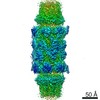 9257C 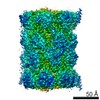 9258C 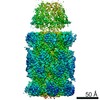 9259C 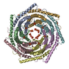 6dfkC 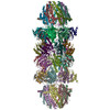 6muvC 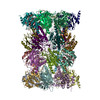 6muwC 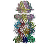 6muxC C: citing same article ( |
|---|---|
| Similar structure data |

-Links
| EMDB pages | EMDB (EBI/PDBe) / EMDataResource |
|---|---|
| Related items in Molecule of the Month |


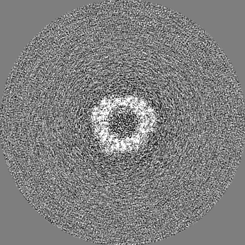
-Map
| File | Download / File: emd_20073.map.gz / Format: CCP4 / Size: 155.3 MB / Type: IMAGE STORED AS FLOATING POINT NUMBER (4 BYTES) | ||||||||||||||||||||||||||||||||||||||||||||||||||||||||||||
|---|---|---|---|---|---|---|---|---|---|---|---|---|---|---|---|---|---|---|---|---|---|---|---|---|---|---|---|---|---|---|---|---|---|---|---|---|---|---|---|---|---|---|---|---|---|---|---|---|---|---|---|---|---|---|---|---|---|---|---|---|---|
| Annotation | Unsharpened map | ||||||||||||||||||||||||||||||||||||||||||||||||||||||||||||
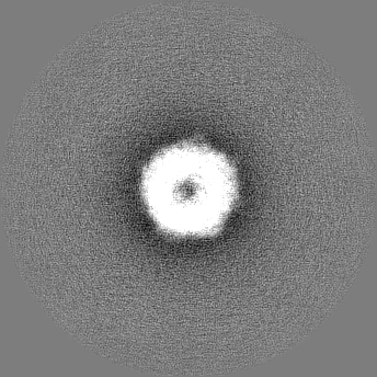
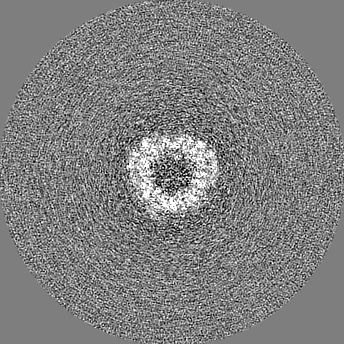
| Projections & slices | Image control
Images are generated by Spider. | ||||||||||||||||||||||||||||||||||||||||||||||||||||||||||||
| Voxel size | X=Y=Z: 1.31 Å | ||||||||||||||||||||||||||||||||||||||||||||||||||||||||||||
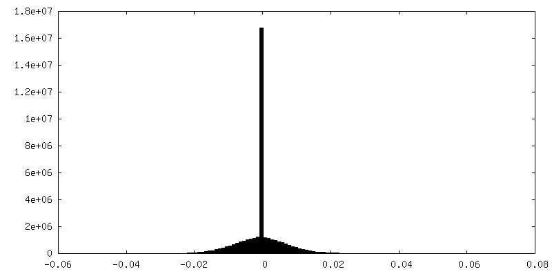
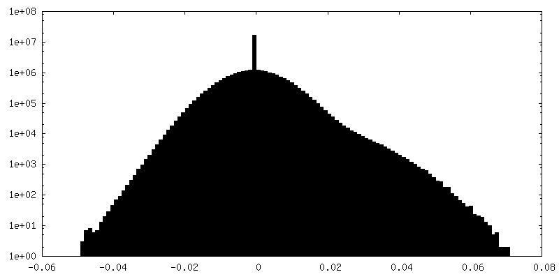
| Density |
| ||||||||||||||||||||||||||||||||||||||||||||||||||||||||||||
| Symmetry | Space group: 1 | ||||||||||||||||||||||||||||||||||||||||||||||||||||||||||||
| Details | EMDB XML:
CCP4 map header:
| ||||||||||||||||||||||||||||||||||||||||||||||||||||||||||||
 Z (Sec.)
 Y (Row.)
 X (Col.)


-Supplemental data
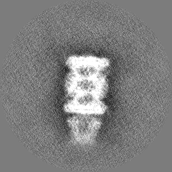
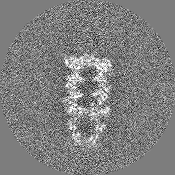
-Half map: Half-map 1
| File | emd_20073_half_map_1.map | ||||||||||||
|---|---|---|---|---|---|---|---|---|---|---|---|---|---|
| Annotation | Half-map 1 | ||||||||||||
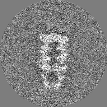
| Projections & Slices |
| ||||||||||||
| Density Histograms |
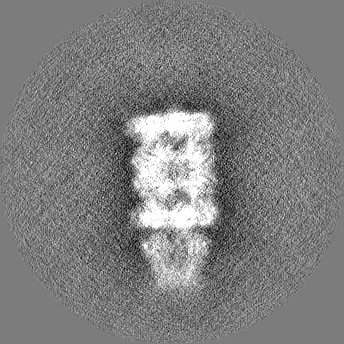

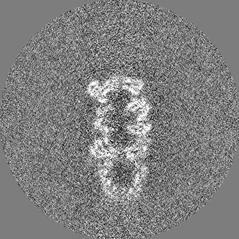
-Half map: Half-map 2
| File | emd_20073_half_map_2.map | ||||||||||||
|---|---|---|---|---|---|---|---|---|---|---|---|---|---|
| Annotation | Half-map 2 | ||||||||||||
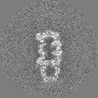
| Projections & Slices |
| ||||||||||||
| Density Histograms |


- Sample components
-Entire : 20S proteasome/PA28 complex.
| Entire | Name: 20S proteasome/PA28 complex. |
|---|---|
| Components |
|
-Supramolecule #1: 20S proteasome/PA28 complex.
| Supramolecule | Name: 20S proteasome/PA28 complex. / type: complex / ID: 1 / Parent: 0 / Macromolecule list: #1-#15 Details: Sample was a mixture of unbound 20S proteasome, 20S proteasome in complex with one PA28 cap, and 20S proteasome in complex with two PA28 caps. |
|---|---|
| Source (natural) | Organism: |
| Molecular weight | Theoretical: 230 KDa |
-Supramolecule #2: Plasmodium falciparum 20S proteasome
| Supramolecule | Name: Plasmodium falciparum 20S proteasome / type: complex / ID: 2 / Parent: 1 / Macromolecule list: #1-#14 |
|---|---|
| Source (natural) | Organism: |
-Supramolecule #3: 11S activator of the Plasmodium falciparum proteasome.
| Supramolecule | Name: 11S activator of the Plasmodium falciparum proteasome. type: complex / ID: 3 / Parent: 1 / Macromolecule list: #15 |
|---|---|
| Source (natural) | Organism: |
| Recombinant expression | Organism:  |
-Experimental details
-Structure determination
| Method | cryo EM |
|---|---|
 Processing Processing | single particle reconstruction |
| Aggregation state | particle |
-Sample preparation
| Concentration | 0.7 mg/mL | |||||||||||||||
|---|---|---|---|---|---|---|---|---|---|---|---|---|---|---|---|---|
| Buffer | pH: 7.4 Component:
| |||||||||||||||
| Grid | Details: unspecified | |||||||||||||||
| Vitrification | Cryogen name: ETHANE / Chamber humidity: 100 % / Chamber temperature: 277.15 K / Instrument: FEI VITROBOT MARK IV / Details: wait time 0sec blot time 2sec blot force -1. |
- Electron microscopy
| Microscope | FEI TALOS ARCTICA |
|---|---|
| Image recording | Film or detector model: GATAN K2 QUANTUM (4k x 4k) / Detector mode: SUPER-RESOLUTION / Digitization - Dimensions - Width: 7676 pixel / Digitization - Dimensions - Height: 7420 pixel / Digitization - Sampling interval: 5.0 µm / Digitization - Frames/image: 1-40 / Number grids imaged: 1 / Number real images: 622 / Average exposure time: 10.0 sec. / Average electron dose: 32.0 e/Å2 |
| Electron beam | Acceleration voltage: 200 kV / Electron source:  FIELD EMISSION GUN FIELD EMISSION GUN |
| Electron optics | C2 aperture diameter: 100.0 µm / Calibrated defocus max: 3.0 µm / Illumination mode: FLOOD BEAM / Imaging mode: BRIGHT FIELD / Cs: 2.7 mm / Nominal defocus min: 1.0 µm / Nominal magnification: 100000 |
| Sample stage | Specimen holder model: FEI TITAN KRIOS AUTOGRID HOLDER / Cooling holder cryogen: NITROGEN |
| Experimental equipment |  Model: Talos Arctica / Image courtesy: FEI Company |

