National Health and Medical Research Council (Australia)
オーストラリア
Australian Research Council
オーストラリア
引用
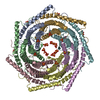
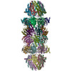
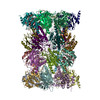
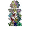
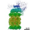
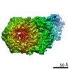
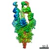
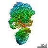
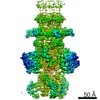
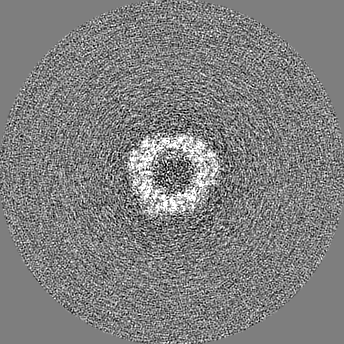
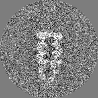
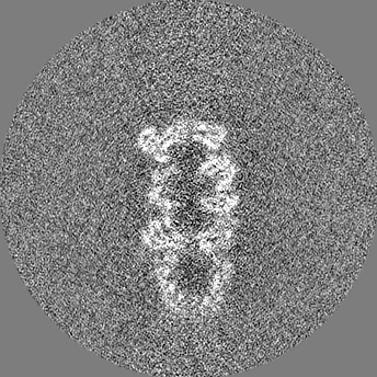
ジャーナル: Nat Microbiol / 年: 2019 タイトル: The structure of the PA28-20S proteasome complex from Plasmodium falciparum and implications for proteostasis. 著者: Stanley C Xie / Riley D Metcalfe / Eric Hanssen / Tuo Yang / David L Gillett / Andrew P Leis / Craig J Morton / Michael J Kuiper / Michael W Parker / Natalie J Spillman / Wilson Wong / ...著者: Stanley C Xie / Riley D Metcalfe / Eric Hanssen / Tuo Yang / David L Gillett / Andrew P Leis / Craig J Morton / Michael J Kuiper / Michael W Parker / Natalie J Spillman / Wilson Wong / Christopher Tsu / Lawrence R Dick / Michael D W Griffin / Leann Tilley / 要旨: The activity of the proteasome 20S catalytic core is regulated by protein complexes that bind to one or both ends. The PA28 regulator stimulates 20S proteasome peptidase activity in vitro, but its ...The activity of the proteasome 20S catalytic core is regulated by protein complexes that bind to one or both ends. The PA28 regulator stimulates 20S proteasome peptidase activity in vitro, but its role in vivo remains unclear. Here, we show that genetic deletion of the PA28 regulator from Plasmodium falciparum (Pf) renders malaria parasites more sensitive to the antimalarial drug dihydroartemisinin, indicating that PA28 may play a role in protection against proteotoxic stress. The crystal structure of PfPA28 reveals a bell-shaped molecule with an inner pore that has a strong segregation of charges. Small-angle X-ray scattering shows that disordered loops, which are not resolved in the crystal structure, extend from the PfPA28 heptamer and surround the pore. Using single particle cryo-electron microscopy, we solved the structure of Pf20S in complex with one and two regulatory PfPA28 caps at resolutions of 3.9 and 3.8 Å, respectively. PfPA28 binds Pf20S asymmetrically, strongly engaging subunits on only one side of the core. PfPA28 undergoes rigid body motions relative to Pf20S. Molecular dynamics simulations support conformational flexibility and a leaky interface. We propose lateral transfer of short peptides through the dynamic interface as a mechanism facilitating the release of proteasome degradation products.
複合体: 11S activator of the Plasmodium falciparum proteasome.
-
超分子 #1: 20S proteasome/PA28 complex.
超分子
名称: 20S proteasome/PA28 complex. / タイプ: complex / ID: 1 / 親要素: 0 / 含まれる分子: #1-#15 詳細: Sample was a mixture of unbound 20S proteasome, 20S proteasome in complex with one PA28 cap, and 20S proteasome in complex with two PA28 caps.
 ムービー
ムービー コントローラー


 データを開く
 基本情報
基本情報 マップデータ
 試料
試料 機能・相同性情報
 Neddylation / Antigen processing: Ubiquitination & Proteasome degradation / proteasome activator complex / Neutrophil degranulation / proteasome core complex / proteasomal ubiquitin-independent protein catabolic process / regulation of G1/S transition of mitotic cell cycle / endopeptidase activator activity /
 データ登録者
 オーストラリア, 2件
  引用
引用
 構造の表示
構造の表示
 ダウンロードとリンク
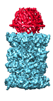 emd_20073.png
 http://ftp.pdbj.org/pub/emdb/structures/EMD-20073


 Z
Z Y
Y X
X


 試料の構成要素
 解析
解析 電子顕微鏡法

