 Movie
Movie Controller

[English] 日本語
 Yorodumi
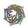
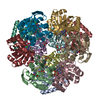
- PDB-6dfk: Crystal structure of the 11S subunit of the Plasmodium falciparum... -
+ Open data

- Basic information
| Entry | Database: PDB / ID: 6dfk | |||||||||||||||
|---|---|---|---|---|---|---|---|---|---|---|---|---|---|---|---|---|
| Title | Crystal structure of the 11S subunit of the Plasmodium falciparum proteasome, PA28 | |||||||||||||||
 Components Components | Subunit of proteaseome activator complex,putative | |||||||||||||||
 Keywords Keywords | PROTEIN BINDING / 11S proteasome subunit / 11S regulatory particle / PA28 / REG / proteasome activator / hydrolase activator | |||||||||||||||
| Function / homology |  Function and homology information Function and homology informationER-Phagosome pathway / Cross-presentation of soluble exogenous antigens (endosomes) / Proteasome assembly / Antigen processing: Ub, ATP-independent proteasomal degradation / proteasome activator complex / endopeptidase activator activity / regulation of G1/S transition of mitotic cell cycle / regulation of proteasomal protein catabolic process / nucleoplasm / cytoplasm Similarity search - Function | |||||||||||||||
| Biological species |  | |||||||||||||||
| Method |  X-RAY DIFFRACTION / SYNCHROTRON / MOLECULAR REPLACEMENT / Resolution: 3.1 Å X-RAY DIFFRACTION / SYNCHROTRON / MOLECULAR REPLACEMENT / Resolution: 3.1 Å | |||||||||||||||
 Authors Authors | Xie, S.C. / Metcalfe, R.D. / Gillett, D.L. / Tilley, L. / Griffin, M.D.W. | |||||||||||||||
| Funding support |  Australia, 4items Australia, 4items
| |||||||||||||||
 Citation Citation | Journal: Nat Microbiol / Year: 2019 Title: The structure of the PA28-20S proteasome complex from Plasmodium falciparum and implications for proteostasis. Authors: Stanley C Xie / Riley D Metcalfe / Eric Hanssen / Tuo Yang / David L Gillett / Andrew P Leis / Craig J Morton / Michael J Kuiper / Michael W Parker / Natalie J Spillman / Wilson Wong / ...Authors: Stanley C Xie / Riley D Metcalfe / Eric Hanssen / Tuo Yang / David L Gillett / Andrew P Leis / Craig J Morton / Michael J Kuiper / Michael W Parker / Natalie J Spillman / Wilson Wong / Christopher Tsu / Lawrence R Dick / Michael D W Griffin / Leann Tilley /  Abstract: The activity of the proteasome 20S catalytic core is regulated by protein complexes that bind to one or both ends. The PA28 regulator stimulates 20S proteasome peptidase activity in vitro, but its ...The activity of the proteasome 20S catalytic core is regulated by protein complexes that bind to one or both ends. The PA28 regulator stimulates 20S proteasome peptidase activity in vitro, but its role in vivo remains unclear. Here, we show that genetic deletion of the PA28 regulator from Plasmodium falciparum (Pf) renders malaria parasites more sensitive to the antimalarial drug dihydroartemisinin, indicating that PA28 may play a role in protection against proteotoxic stress. The crystal structure of PfPA28 reveals a bell-shaped molecule with an inner pore that has a strong segregation of charges. Small-angle X-ray scattering shows that disordered loops, which are not resolved in the crystal structure, extend from the PfPA28 heptamer and surround the pore. Using single particle cryo-electron microscopy, we solved the structure of Pf20S in complex with one and two regulatory PfPA28 caps at resolutions of 3.9 and 3.8 Å, respectively. PfPA28 binds Pf20S asymmetrically, strongly engaging subunits on only one side of the core. PfPA28 undergoes rigid body motions relative to Pf20S. Molecular dynamics simulations support conformational flexibility and a leaky interface. We propose lateral transfer of short peptides through the dynamic interface as a mechanism facilitating the release of proteasome degradation products. | |||||||||||||||
| History |
|
- Structure visualization
| Structure viewer | Molecule: MolmilJmol/JSmol |
|---|
- Downloads & links
-Download
| PDBx/mmCIF format | 6dfk.cif.gz | 1.3 MB | Display | PDBx/mmCIF format |
|---|---|---|---|---|
| PDB format | pdb6dfk.ent.gz | 1.1 MB | Display | PDB format |
| PDBx/mmJSON format | 6dfk.json.gz | Tree view | PDBx/mmJSON format | |
| Others |  Other downloads Other downloads |
-Validation report
| Arichive directory | https://data.pdbj.org/pub/pdb/validation_reports/df/6dfkftp://data.pdbj.org/pub/pdb/validation_reports/df/6dfk | HTTPS FTP |
|---|
-Related structure data
| Related structure data | 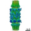 9257C 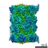 9258C 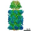 9259C 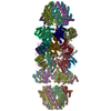 6muvC  6muwC 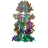 6muxC  1avoS S: Starting model for refinement C: citing same article ( |
|---|---|
| Similar structure data |


-Links
 PDBj
PDBj

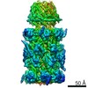
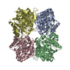
- Assembly
| Deposited unit | 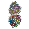
| ||||||||
|---|---|---|---|---|---|---|---|---|---|
| 1 | 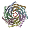
| ||||||||
| 2 | 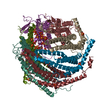
| ||||||||
| Unit cell |
|
-Components
| #1: Protein | Mass: 33235.824 Da / Num. of mol.: 14 Source method: isolated from a genetically manipulated source Source: (gene. exp.) Gene: PF3D7_0907700 / Production host:  #2: Chemical | ChemComp-SO4 /   Mass: 96.063 Da / Num. of mol.: 60 / Source method: obtained synthetically / Formula: SO4 Mass: 96.063 Da / Num. of mol.: 60 / Source method: obtained synthetically / Formula: SO4 |
|---|
-Experimental details
-Experiment
| Experiment | Method: X-RAY DIFFRACTION / Number of used crystals: 1 |
|---|
- Sample preparation
| Crystal | Density Matthews: 3.44 Å3/Da / Density % sol: 64.2 % |
|---|---|
| Crystal grow | Temperature: 293 K / Method: vapor diffusion, sitting drop / pH: 6.5 / Details: 20 mM HEPES, 2 M ammonium sulfate |
-Data collection
| Diffraction | Mean temperature: 100 K |
|---|---|
| Diffraction source | Source: SYNCHROTRON / Site: Australian Synchrotron / Beamline: MX2 / Wavelength: 0.9537 Å |
| Detector | Type: DECTRIS EIGER X 16M / Detector: PIXEL / Date: Jun 14, 2017 |
| Radiation | Protocol: SINGLE WAVELENGTH / Monochromatic (M) / Laue (L): M / Scattering type: x-ray |
| Radiation wavelength | Wavelength: 0.9537 Å / Relative weight: 1 |
| Reflection | Resolution: 3.1→48.89 Å / Num. obs: 116975 / % possible obs: 100 % / Redundancy: 10.4 % / Biso Wilson estimate: 60.6 Å2 / CC1/2: 0.996 / Rpim(I) all: 0.131 / Rrim(I) all: 0.305 / Net I/σ(I): 7.5 |
| Reflection shell | Resolution: 3.1→3.15 Å / Redundancy: 10.7 % / Mean I/σ(I) obs: 1.2 / Num. unique obs: 5711 / CC1/2: 0.513 / % possible all: 100 |
- Processing
| Software |
| |||||||||||||||||||||||||||||||||||||||||||||||||||||||||||||||||||||||||||||||||||||||||||||||||||||||||||||||||||||||||||||||||||||||||||||||||||||||||||||||||||||||||||||||||||||||||||||||||||||||||||||||||||||||||||||||||||||||||||||||||||||||||||||||||||||||||||||||||||||||||||||||||||||||||||||||||||||||||||||||||||||||||||||||||||||||||||||||||||||||||||||||||||||||
|---|---|---|---|---|---|---|---|---|---|---|---|---|---|---|---|---|---|---|---|---|---|---|---|---|---|---|---|---|---|---|---|---|---|---|---|---|---|---|---|---|---|---|---|---|---|---|---|---|---|---|---|---|---|---|---|---|---|---|---|---|---|---|---|---|---|---|---|---|---|---|---|---|---|---|---|---|---|---|---|---|---|---|---|---|---|---|---|---|---|---|---|---|---|---|---|---|---|---|---|---|---|---|---|---|---|---|---|---|---|---|---|---|---|---|---|---|---|---|---|---|---|---|---|---|---|---|---|---|---|---|---|---|---|---|---|---|---|---|---|---|---|---|---|---|---|---|---|---|---|---|---|---|---|---|---|---|---|---|---|---|---|---|---|---|---|---|---|---|---|---|---|---|---|---|---|---|---|---|---|---|---|---|---|---|---|---|---|---|---|---|---|---|---|---|---|---|---|---|---|---|---|---|---|---|---|---|---|---|---|---|---|---|---|---|---|---|---|---|---|---|---|---|---|---|---|---|---|---|---|---|---|---|---|---|---|---|---|---|---|---|---|---|---|---|---|---|---|---|---|---|---|---|---|---|---|---|---|---|---|---|---|---|---|---|---|---|---|---|---|---|---|---|---|---|---|---|---|---|---|---|---|---|---|---|---|---|---|---|---|---|---|---|---|---|---|---|---|---|---|---|---|---|---|---|---|---|---|---|---|---|---|---|---|---|---|---|---|---|---|---|---|---|---|---|---|---|---|---|---|---|---|---|---|---|---|---|---|---|---|---|---|---|---|---|---|---|---|---|---|---|---|---|---|---|---|---|---|---|---|---|---|---|---|---|---|---|---|---|---|---|---|---|---|---|---|---|
| Refinement | Method to determine structure: MOLECULAR REPLACEMENT Starting model: 1AVO Resolution: 3.1→48.89 Å / SU ML: 0.34 / Cross valid method: FREE R-VALUE / σ(F): 1.01 / Phase error: 22.42
| |||||||||||||||||||||||||||||||||||||||||||||||||||||||||||||||||||||||||||||||||||||||||||||||||||||||||||||||||||||||||||||||||||||||||||||||||||||||||||||||||||||||||||||||||||||||||||||||||||||||||||||||||||||||||||||||||||||||||||||||||||||||||||||||||||||||||||||||||||||||||||||||||||||||||||||||||||||||||||||||||||||||||||||||||||||||||||||||||||||||||||||||||||||||
| Solvent computation | Shrinkage radii: 0.9 Å / VDW probe radii: 1.11 Å | |||||||||||||||||||||||||||||||||||||||||||||||||||||||||||||||||||||||||||||||||||||||||||||||||||||||||||||||||||||||||||||||||||||||||||||||||||||||||||||||||||||||||||||||||||||||||||||||||||||||||||||||||||||||||||||||||||||||||||||||||||||||||||||||||||||||||||||||||||||||||||||||||||||||||||||||||||||||||||||||||||||||||||||||||||||||||||||||||||||||||||||||||||||||
| Refinement step | Cycle: LAST / Resolution: 3.1→48.89 Å
| |||||||||||||||||||||||||||||||||||||||||||||||||||||||||||||||||||||||||||||||||||||||||||||||||||||||||||||||||||||||||||||||||||||||||||||||||||||||||||||||||||||||||||||||||||||||||||||||||||||||||||||||||||||||||||||||||||||||||||||||||||||||||||||||||||||||||||||||||||||||||||||||||||||||||||||||||||||||||||||||||||||||||||||||||||||||||||||||||||||||||||||||||||||||
| Refine LS restraints |
| |||||||||||||||||||||||||||||||||||||||||||||||||||||||||||||||||||||||||||||||||||||||||||||||||||||||||||||||||||||||||||||||||||||||||||||||||||||||||||||||||||||||||||||||||||||||||||||||||||||||||||||||||||||||||||||||||||||||||||||||||||||||||||||||||||||||||||||||||||||||||||||||||||||||||||||||||||||||||||||||||||||||||||||||||||||||||||||||||||||||||||||||||||||||
| LS refinement shell |
| |||||||||||||||||||||||||||||||||||||||||||||||||||||||||||||||||||||||||||||||||||||||||||||||||||||||||||||||||||||||||||||||||||||||||||||||||||||||||||||||||||||||||||||||||||||||||||||||||||||||||||||||||||||||||||||||||||||||||||||||||||||||||||||||||||||||||||||||||||||||||||||||||||||||||||||||||||||||||||||||||||||||||||||||||||||||||||||||||||||||||||||||||||||||
| Refinement TLS params. | Method: refined / Refine-ID: X-RAY DIFFRACTION
| |||||||||||||||||||||||||||||||||||||||||||||||||||||||||||||||||||||||||||||||||||||||||||||||||||||||||||||||||||||||||||||||||||||||||||||||||||||||||||||||||||||||||||||||||||||||||||||||||||||||||||||||||||||||||||||||||||||||||||||||||||||||||||||||||||||||||||||||||||||||||||||||||||||||||||||||||||||||||||||||||||||||||||||||||||||||||||||||||||||||||||||||||||||||
| Refinement TLS group |
|
