Movie
Movie Controller


+ Open data

- Basic information
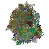
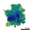
| Entry | Database: EMDB / ID: EMD-5189 | |||||||||
|---|---|---|---|---|---|---|---|---|---|---|
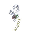
| Title | tmRNA-SmpB: a journey to the center of the bacterial ribosome | |||||||||
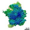
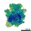
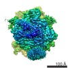
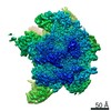
 Map data Map data | tmRNA-SmpB complex translocated in the bacterial ribosome P site | |||||||||
 Sample Sample |
| |||||||||
 Keywords Keywords | TMRNA / SMPB / RIBOSOME / TRANS-TRANSLATION | |||||||||
| Function / homology |  Function and homology information Function and homology informationtrans-translation-dependent protein tagging / trans-translation / rRNA binding / cytosol Similarity search - Function | |||||||||
| Biological species |   Thermus thermophilus (bacteria) Thermus thermophilus (bacteria) | |||||||||
| Method | single particle reconstruction / cryo EM / Resolution: 13.0 Å | |||||||||
 Authors Authors | Weis F / Bron P / Giudice E / Rolland JP / Thomas D / Felden B / Gillet R | |||||||||
 Citation Citation | Journal: EMBO J / Year: 2010 Title: tmRNA-SmpB: a journey to the centre of the bacterial ribosome. Authors: Félix Weis / Patrick Bron / Emmanuel Giudice / Jean-Paul Rolland / Daniel Thomas / Brice Felden / Reynald Gillet /  Abstract: Ribosomes mediate protein synthesis by decoding the information carried by messenger RNAs (mRNAs) and catalysing peptide bond formation between amino acids. When bacterial ribosomes stall on ...Ribosomes mediate protein synthesis by decoding the information carried by messenger RNAs (mRNAs) and catalysing peptide bond formation between amino acids. When bacterial ribosomes stall on incomplete messages, the trans-translation quality control mechanism is activated by the transfer-messenger RNA bound to small protein B (tmRNA-SmpB ribonucleoprotein complex). Trans-translation liberates the stalled ribosomes and triggers degradation of the incomplete proteins. Here, we present the cryo-electron microscopy structures of tmRNA-SmpB accommodated or translocated into stalled ribosomes. Two atomic models for each state are proposed. This study reveals how tmRNA-SmpB crosses the ribosome and how, as the problematic mRNA is ejected, the tmRNA resume codon is placed onto the ribosomal decoding site by new contacts between SmpB and the nucleotides upstream of the tag-encoding sequence. This provides a structural basis for the transit of the large tmRNA-SmpB complex through the ribosome and for the means by which the tmRNA internal frame is set for translation to resume. | |||||||||
| History |
|
- Structure visualization
| Movie |
Movie viewer |
|---|---|
| Structure viewer | EM map: SurfViewMolmilJmol/JSmol |
| Supplemental images |

- Downloads & links
-EMDB archive
| Map data | emd_5189.map.gz | 1.4 MB | EMDB map data format | |
|---|---|---|---|---|
| Header (meta data) | emd-5189-v30.xmlemd-5189.xml | 11.6 KB 11.6 KB | Display Display | EMDB header |
| Images | 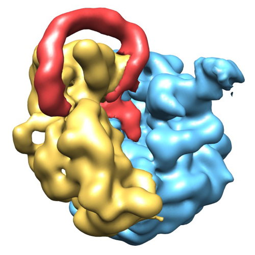 emd_5189_1.png emd_5189_1.png | 273.9 KB | ||
| Archive directory |  http://ftp.pdbj.org/pub/emdb/structures/EMD-5189ftp://ftp.pdbj.org/pub/emdb/structures/EMD-5189 http://ftp.pdbj.org/pub/emdb/structures/EMD-5189ftp://ftp.pdbj.org/pub/emdb/structures/EMD-5189 | HTTPS FTP |
-Related structure data
| Related structure data |  3iyrMC  5188C  3iyqC M: atomic model generated by this map C: citing same article ( |
|---|---|
| Similar structure data |

-Links
| EMDB pages | EMDB (EBI/PDBe) / EMDataResource |
|---|---|
| Related items in Molecule of the Month |


-Map
| File | Download / File: emd_5189.map.gz / Format: CCP4 / Size: 7.8 MB / Type: IMAGE STORED AS FLOATING POINT NUMBER (4 BYTES) | ||||||||||||||||||||||||||||||||||||||||||||||||||||||||||||||||||||
|---|---|---|---|---|---|---|---|---|---|---|---|---|---|---|---|---|---|---|---|---|---|---|---|---|---|---|---|---|---|---|---|---|---|---|---|---|---|---|---|---|---|---|---|---|---|---|---|---|---|---|---|---|---|---|---|---|---|---|---|---|---|---|---|---|---|---|---|---|---|
| Annotation | tmRNA-SmpB complex translocated in the bacterial ribosome P site | ||||||||||||||||||||||||||||||||||||||||||||||||||||||||||||||||||||
| Projections & slices | Image control
Images are generated by Spider. | ||||||||||||||||||||||||||||||||||||||||||||||||||||||||||||||||||||
| Voxel size | X=Y=Z: 3.28 Å | ||||||||||||||||||||||||||||||||||||||||||||||||||||||||||||||||||||
| Density |
| ||||||||||||||||||||||||||||||||||||||||||||||||||||||||||||||||||||
| Symmetry | Space group: 1 | ||||||||||||||||||||||||||||||||||||||||||||||||||||||||||||||||||||
| Details | EMDB XML:
CCP4 map header:
| ||||||||||||||||||||||||||||||||||||||||||||||||||||||||||||||||||||
 Z (Sec.)
 Y (Row.)
 X (Col.)


-Supplemental data
- Sample components
-Entire : Thermus thermophilus 70S ribosome
| Entire | Name: Thermus thermophilus 70S ribosome |
|---|---|
| Components |
|
-Supramolecule #1000: Thermus thermophilus 70S ribosome
| Supramolecule | Name: Thermus thermophilus 70S ribosome / type: sample / ID: 1000 / Number unique components: 5 |
|---|---|
| Molecular weight | Theoretical: 2.3 MDa |
-Supramolecule #1: 70S ribosome
| Supramolecule | Name: 70S ribosome / type: complex / ID: 1 / Recombinant expression: No / Database: NCBI / Ribosome-details: ribosome-prokaryote: ALL |
|---|---|
| Source (natural) | Organism: Thermus thermophilus (bacteria) |
| Molecular weight | Theoretical: 2.3 MDa |
-Experimental details
-Structure determination
| Method | cryo EM |
|---|---|
 Processing Processing | single particle reconstruction |
| Aggregation state | particle |
-Sample preparation
| Buffer | pH: 7.5 Details: 5mM Hepes-KOH (pH 7.5), 10mM NH4Cl, 10mM MgOAc, 50mM KCl, 0.1mM EDTA and 6mM BetaME |
|---|---|
| Grid | Details: Quantifoil holey-carbon grids previously glow-discharged |
| Vitrification | Cryogen name: ETHANE / Chamber humidity: 100 % / Chamber temperature: 100 K / Instrument: OTHER / Details: Vitrification instrument: vitrobot / Method: Blot for 5 seconds before plunging |
- Electron microscopy
| Microscope | JEOL 2200FS |
|---|---|
| Temperature | Average: 95 K |
| Specialist optics | Energy filter - Name: JEOL / Energy filter - Lower energy threshold: 0.0 eV / Energy filter - Upper energy threshold: 20.0 eV |
| Date | Jul 1, 2009 |
| Image recording | Category: FILM / Film or detector model: KODAK SO-163 FILM / Digitization - Scanner: NIKON SUPER COOLSCAN 9000 / Digitization - Sampling interval: 7.5 µm / Number real images: 163 / Bits/pixel: 8 |
| Electron beam | Acceleration voltage: 200 kV / Electron source:  FIELD EMISSION GUN FIELD EMISSION GUN |
| Electron optics | Calibrated magnification: 45700 / Illumination mode: FLOOD BEAM / Imaging mode: BRIGHT FIELD / Cs: 2 mm / Nominal defocus max: 2.2 µm / Nominal defocus min: 0.9 µm / Nominal magnification: 50000 |
| Sample stage | Specimen holder: Eucentric / Specimen holder model: GATAN LIQUID NITROGEN |
-Image processing
| CTF correction | Details: Each particle |
|---|---|
| Final reconstruction | Resolution.type: BY AUTHOR / Resolution: 13.0 Å / Resolution method: FSC 0.5 CUT-OFF / Software - Name: IMAGIC-V / Number images used: 70761 |
-Atomic model buiding 1
| Initial model | PDB ID: Chain - #0 - Chain ID: A / Chain - #1 - Chain ID: B |
|---|---|
| Software | Name: UCSF-Chimera, mdff |
| Details | Protocol: Rigid Body and flexible fitting. The domains were initially separately fitted by manual docking and then further optimized using mdff. |
| Refinement | Space: REAL / Protocol: FLEXIBLE FIT / Target criteria: Cross-Correlation |
| Output model | PDB-3iyr: |

-Atomic model buiding 2
| Initial model | PDB ID: Chain - #0 - Chain ID: A / Chain - #1 - Chain ID: B |
|---|---|
| Software | Name: UCSF-Chimera, mdff |
| Details | Protocol: Rigid Body and flexible fitting. each subdomain (H2b-c, H5, pk1, pk2, pk3, pk4) were initially separately fitted by manual docking and then further optimized using mdff. |
| Refinement | Space: REAL / Protocol: FLEXIBLE FIT / Target criteria: Cross-Correlation |
| Output model | PDB-3iyr: |