 ムービー
ムービー コントローラー
 構造ビューア
 EMN検索について


-検索条件
-検索結果
検索 (著者・登録者: burgess & sa)の結果75件中、1から50件目までを表示しています
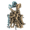
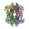
EMDB-17377: 
Structure of human SIT1 (focussed map / refinement)

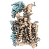
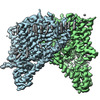
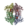
EMDB-17378:
Structure of human SIT1:ACE2 complex (open PD conformation)

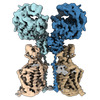
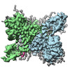
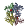
EMDB-17379:
Structure of human SIT1:ACE2 complex (closed PD conformation)
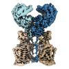
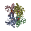
EMDB-17380:
Structure of human SIT1 bound to L-pipecolate (focussed map / refinement)
EMDB-17381:
Structure of human SIT1:ACE2 complex (open PD conformation) bound to L-pipecolate
EMDB-17382:
Structure of human SIT1:ACE2 complex (closed PD conformation) bound to L-pipecolate

PDB-8p2w:
Structure of human SIT1 (focussed map / refinement)

PDB-8p2x:
Structure of human SIT1:ACE2 complex (open PD conformation)

PDB-8p2y:
Structure of human SIT1:ACE2 complex (closed PD conformation)

PDB-8p2z:
Structure of human SIT1 bound to L-pipecolate (focussed map / refinement)
PDB-8p30:
Structure of human SIT1:ACE2 complex (open PD conformation) bound to L-pipecolate

PDB-8p31:
Structure of human SIT1:ACE2 complex (closed PD conformation) bound to L-pipecolate
EMDB-17197:
Human TPC2 in Complex with Antagonist (S)-SG-094
EMDB-19108:
Human TPC2 in Complex withAntagonist (R)-SG-094

PDB-8ouo:
Human TPC2 in Complex with Antagonist (S)-SG-094

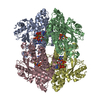
EMDB-18729:
Cryo-EM structure of tetrameric human SAMHD1 with dApNHpp

EMDB-18730:
Cryo-EM structure of tetrameric human SAMHD1 State I - Tense

EMDB-18731:
Cryo-EM structure of tetrameric human SAMHD1 State II - Hemi-relaxed

EMDB-18732:
Cryo-EM structure of tetrameric human SAMHD1 State III - Relaxed

EMDB-18733:
Cryo-EM structure of tetrameric human SAMHD1 State IV - Depleted relaxed

EMDB-18734:
Cryo-EM structure of tetrameric human SAMHD1 State V - Depleted relaxed
PDB-8qxj:
Cryo-EM structure of tetrameric human SAMHD1 with dApNHpp
PDB-8qxk:
Cryo-EM structure of tetrameric human SAMHD1 State I - Tense
PDB-8qxl:
Cryo-EM structure of tetrameric human SAMHD1 State II - Hemi-relaxed
PDB-8qxm:
Cryo-EM structure of tetrameric human SAMHD1 State III - Relaxed
PDB-8qxn:
Cryo-EM structure of tetrameric human SAMHD1 State IV - Depleted relaxed
PDB-8qxo:
Cryo-EM structure of tetrameric human SAMHD1 State V - Depleted relaxed
EMDB-16110:
Human Urea Transporter UT-A (N-Terminal Domain Model)

EMDB-16111:
Map of Human Urea Transporter UT-A Collected with 0 and 30 Degree Tilts
EMDB-16112:
Human Urea Transporter UT-B/UT1 in Complex with Inhibitor UTBinh-14
PDB-8blo:
Human Urea Transporter UT-A (N-Terminal Domain Model)
PDB-8blp:
Human Urea Transporter UT-B/UT1 in Complex with Inhibitor UTBinh-14

EMDB-18301:
In-tissue cryo electron tomograms of App^NL-G-F amyloid plaques
EMDB-16018:
Sarkosyl-extracted AppNL-G-F Abeta42 fibril structure
EMDB-16019:
Sarkosyl-extracted AppNL-G-F Abeta42 fibril structure (Methoxy-X04-labelled mice)

PDB-8bfa:
Sarkosyl-extracted AppNL-G-F Abeta42 fibril structure

PDB-8bfb:
Sarkosyl-extracted AppNL-G-F Abeta42 fibril structure (Methoxy-X04-labelled mice)

EMDB-13036:
Cryo-EM structure of the human TRPA1 ion channel in complex with the antagonist 3-60, conformation 2

EMDB-13037:
Cryo-EM structure of the human TRPA1 ion channel in complex with the antagonist 3-60, conformation 1

PDB-7or0:
Cryo-EM structure of the human TRPA1 ion channel in complex with the antagonist 3-60, conformation 2

PDB-7or1:
Cryo-EM structure of the human TRPA1 ion channel in complex with the antagonist 3-60, conformation 1

EMDB-13416:
Human voltage-gated potassium channel Kv3.1 (apo condition)

EMDB-13417:
Human voltage-gated potassium channel Kv3.1 (with Zn)

EMDB-13418:
Human voltage-gated potassium channel Kv3.1 in dimeric state (with Zn)

EMDB-13419:
Human voltage-gated potassium channel Kv3.1 (with EDTA)

PDB-7phh:
Human voltage-gated potassium channel Kv3.1 (apo condition)

PDB-7phi:
Human voltage-gated potassium channel Kv3.1 (with Zn)

PDB-7phk:
Human voltage-gated potassium channel Kv3.1 in dimeric state (with Zn)

PDB-7phl:
Human voltage-gated potassium channel Kv3.1 (with EDTA)

EMDB-11804:
Structure of Wild-Type Human Potassium Chloride Transporter KCC3 in NaCl (LMNG/CHS)
ページ:
 wwPDBはEMDBデータモデルのバージョン3へ移行します
