 Movie
Movie Controller

[English] 日本語
 Yorodumi
- EMDB-16019: Sarkosyl-extracted AppNL-G-F Abeta42 fibril structure (Methoxy-X0... -
+ Open data

- Basic information
| Entry |  | |||||||||
|---|---|---|---|---|---|---|---|---|---|---|
| Title | Sarkosyl-extracted AppNL-G-F Abeta42 fibril structure (Methoxy-X04-labelled mice) | |||||||||
 Map data Map data | CryoEM map for extracted AppNLGF Abeta42 fibril after MOX04 labelling | |||||||||
 Sample Sample |
| |||||||||
 Keywords Keywords | Amyloid / fibril / helical / cross-beta / beta amyloid / PROTEIN FIBRIL / ex vivo / arctic mutant / alzheimers disease | |||||||||
| Function / homology |  Function and homology information Function and homology informationamyloid-beta complex / growth cone lamellipodium / cellular response to norepinephrine stimulus / collateral sprouting in absence of injury / growth cone filopodium / microglia development / hippocampal neuron apoptotic process / Formyl peptide receptors bind formyl peptides and many other ligands / regulation of Wnt signaling pathway / axo-dendritic transport ...amyloid-beta complex / growth cone lamellipodium / cellular response to norepinephrine stimulus / collateral sprouting in absence of injury / growth cone filopodium / microglia development / hippocampal neuron apoptotic process / Formyl peptide receptors bind formyl peptides and many other ligands / regulation of Wnt signaling pathway / axo-dendritic transport / axon midline choice point recognition / regulation of synapse structure or activity / astrocyte activation involved in immune response / NMDA selective glutamate receptor signaling pathway / regulation of spontaneous synaptic transmission / mating behavior / growth factor receptor binding / peptidase activator activity / Insertion of tail-anchored proteins into the endoplasmic reticulum membrane / positive regulation of amyloid fibril formation / Golgi-associated vesicle / PTB domain binding / astrocyte projection / Lysosome Vesicle Biogenesis / Deregulated CDK5 triggers multiple neurodegenerative pathways in Alzheimer's disease models / neuron remodeling / nuclear envelope lumen / dendrite development / regulation of multicellular organism growth / TRAF6 mediated NF-kB activation / positive regulation of protein metabolic process / signaling receptor activator activity / negative regulation of long-term synaptic potentiation / transition metal ion binding / Advanced glycosylation endproduct receptor signaling / The NLRP3 inflammasome / modulation of excitatory postsynaptic potential / main axon / intracellular copper ion homeostasis / ECM proteoglycans / response to insulin-like growth factor stimulus / positive regulation of T cell migration / regulation of presynapse assembly / Notch signaling pathway / neuronal dense core vesicle / swimming behavior / Purinergic signaling in leishmaniasis infection / positive regulation of mitotic cell cycle / adult locomotory behavior / cellular response to manganese ion / positive regulation of chemokine production / extracellular matrix organization / positive regulation of calcium-mediated signaling / axonogenesis / neuron projection maintenance / clathrin-coated pit / Mitochondrial protein degradation / ionotropic glutamate receptor signaling pathway / astrocyte activation / platelet alpha granule lumen / response to interleukin-1 / regulation of neuron apoptotic process / positive regulation of glycolytic process / cellular response to cAMP / cellular response to copper ion / endosome lumen / trans-Golgi network membrane / positive regulation of interleukin-1 beta production / learning / protein serine/threonine kinase binding / dendritic shaft / positive regulation of long-term synaptic potentiation / central nervous system development / Post-translational protein phosphorylation / serine-type endopeptidase inhibitor activity / regulation of long-term neuronal synaptic plasticity / locomotory behavior / microglial cell activation / cellular response to nerve growth factor stimulus / visual learning / positive regulation of non-canonical NF-kappaB signal transduction / TAK1-dependent IKK and NF-kappa-B activation / synapse organization / positive regulation of JNK cascade / recycling endosome / response to lead ion / positive regulation of interleukin-6 production / Golgi lumen / cognition / Regulation of Insulin-like Growth Factor (IGF) transport and uptake by Insulin-like Growth Factor Binding Proteins (IGFBPs) / cellular response to amyloid-beta / neuron projection development / endocytosis / calcium ion transport / positive regulation of inflammatory response / positive regulation of tumor necrosis factor production / Platelet degranulation / heparin binding / regulation of translation / regulation of gene expression Similarity search - Function | |||||||||
| Biological species |  | |||||||||
| Method | helical reconstruction / cryo EM / Resolution: 3.2 Å | |||||||||
 Authors Authors | Wilkinson M / Leistner C / Burgess A / Goodfellow S / Deuchars S / Ranson NA / Radford SE / Frank RAW | |||||||||
| Funding support |  United Kingdom, 2 items United Kingdom, 2 items
| |||||||||
 Citation Citation | Journal: Nat Commun / Year: 2023 Title: The in-tissue molecular architecture of β-amyloid pathology in the mammalian brain. Authors: Conny Leistner / Martin Wilkinson / Ailidh Burgess / Megan Lovatt / Stanley Goodbody / Yong Xu / Susan Deuchars / Sheena E Radford / Neil A Ranson / René A W Frank / Abstract: Amyloid plaques composed of Aβ fibrils are a hallmark of Alzheimer's disease (AD). However, the molecular architecture of amyloid plaques in the context of fresh mammalian brain tissue is unknown. ...Amyloid plaques composed of Aβ fibrils are a hallmark of Alzheimer's disease (AD). However, the molecular architecture of amyloid plaques in the context of fresh mammalian brain tissue is unknown. Here, using cryogenic correlated light and electron tomography we report the in situ molecular architecture of Aβ fibrils in the App familial AD mouse model containing the Arctic mutation and an atomic model of ex vivo purified Arctic Aβ fibrils. We show that in-tissue Aβ fibrils are arranged in a lattice or parallel bundles, and are interdigitated by subcellular compartments, extracellular vesicles, extracellular droplets and extracellular multilamellar bodies. The Arctic Aβ fibril differs significantly from an earlier App fibril structure, indicating a striking effect of the Arctic mutation. These structural data also revealed an ensemble of additional fibrillar species, including thin protofilament-like rods and branched fibrils. Together, these results provide a structural model for the dense network architecture that characterises β-amyloid plaque pathology. | |||||||||
| History |
|
- Structure visualization
| Supplemental images |
|---|

- Downloads & links
-EMDB archive
| Map data | emd_16019.map.gz | 5.7 MB | EMDB map data format | |
|---|---|---|---|---|
| Header (meta data) | emd-16019-v30.xmlemd-16019.xml | 18.2 KB 18.2 KB | Display Display | EMDB header |
| Images | 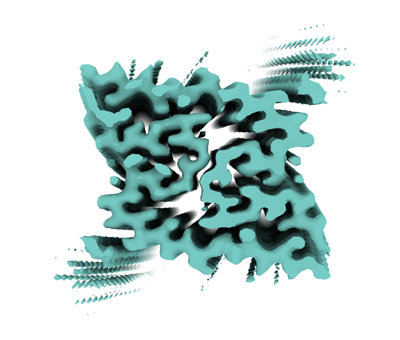 emd_16019.png emd_16019.png | 91.5 KB | ||
| Filedesc metadata | emd-16019.cif.gz | 5.8 KB | ||
| Others | emd_16019_half_map_1.map.gzemd_16019_half_map_2.map.gz | 56.6 MB 56.7 MB | ||
| Archive directory |  http://ftp.pdbj.org/pub/emdb/structures/EMD-16019ftp://ftp.pdbj.org/pub/emdb/structures/EMD-16019 http://ftp.pdbj.org/pub/emdb/structures/EMD-16019ftp://ftp.pdbj.org/pub/emdb/structures/EMD-16019 | HTTPS FTP |
-Related structure data
| Related structure data |  8bfbMC  8bfaC M: atomic model generated by this map C: citing same article ( |
|---|---|
| Similar structure data |


-Links
| EMDB pages | EMDB (EBI/PDBe) / EMDataResource |
|---|---|
| Related items in Molecule of the Month |


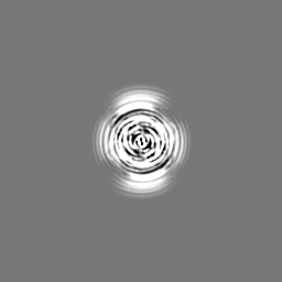
-Map
| File | Download / File: emd_16019.map.gz / Format: CCP4 / Size: 64 MB / Type: IMAGE STORED AS FLOATING POINT NUMBER (4 BYTES) | ||||||||||||||||||||||||||||||||||||
|---|---|---|---|---|---|---|---|---|---|---|---|---|---|---|---|---|---|---|---|---|---|---|---|---|---|---|---|---|---|---|---|---|---|---|---|---|---|
| Annotation | CryoEM map for extracted AppNLGF Abeta42 fibril after MOX04 labelling | ||||||||||||||||||||||||||||||||||||
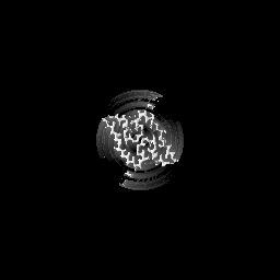
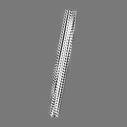
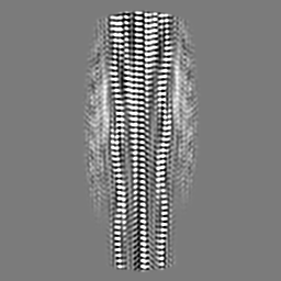
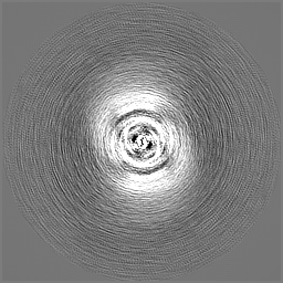
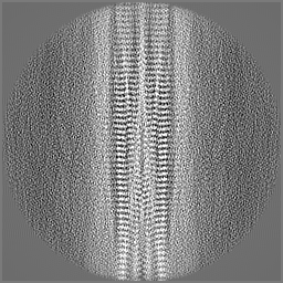
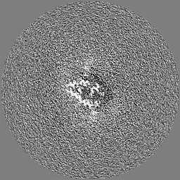
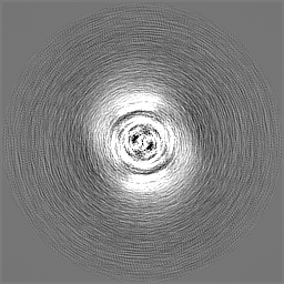
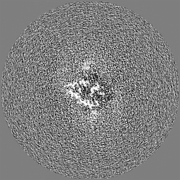
| Projections & slices | Image control
Images are generated by Spider. | ||||||||||||||||||||||||||||||||||||
| Voxel size | X=Y=Z: 0.94 Å | ||||||||||||||||||||||||||||||||||||
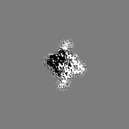
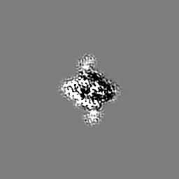
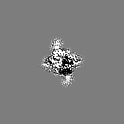
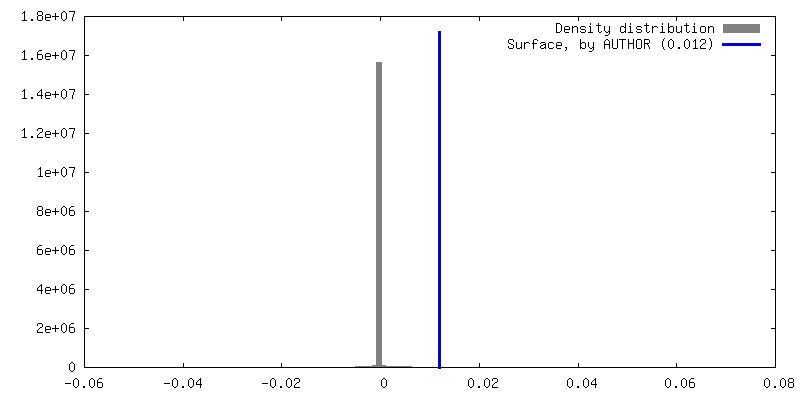
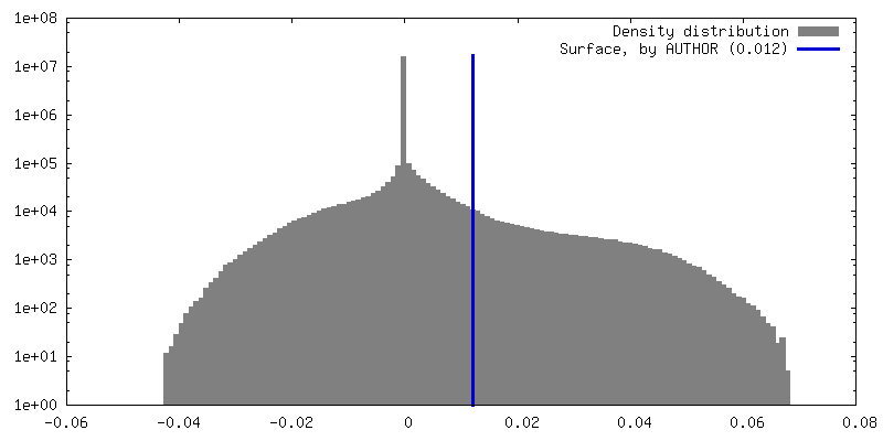
| Density |
| ||||||||||||||||||||||||||||||||||||
| Symmetry | Space group: 1 | ||||||||||||||||||||||||||||||||||||
| Details | EMDB XML:
|
 Z (Sec.)
 Y (Row.)
 X (Col.)


-Supplemental data
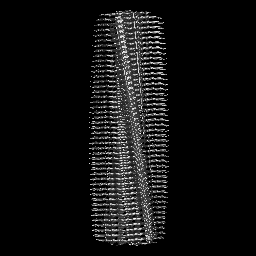
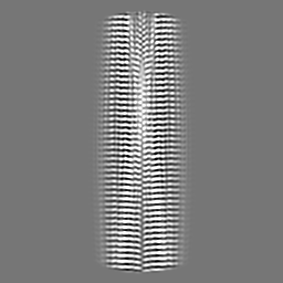
-Half map: halfmap2
| File | emd_16019_half_map_1.map | ||||||||||||
|---|---|---|---|---|---|---|---|---|---|---|---|---|---|
| Annotation | halfmap2 | ||||||||||||
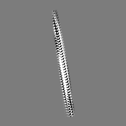
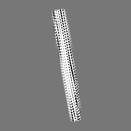
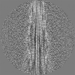
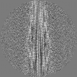
| Projections & Slices |
| ||||||||||||
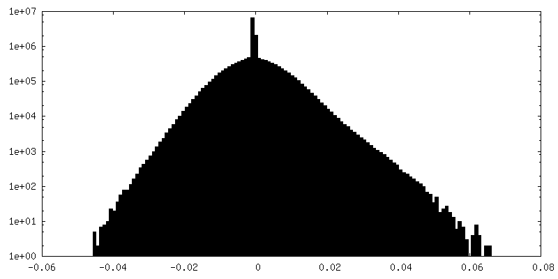
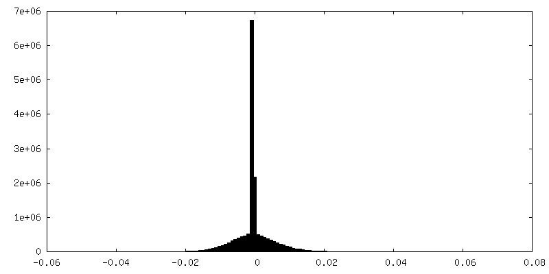
| Density Histograms |


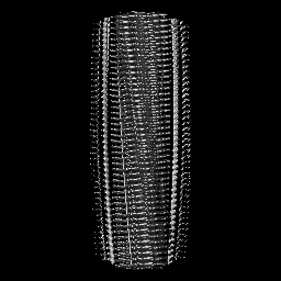
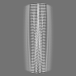
-Half map: halfmap1
| File | emd_16019_half_map_2.map | ||||||||||||
|---|---|---|---|---|---|---|---|---|---|---|---|---|---|
| Annotation | halfmap1 | ||||||||||||
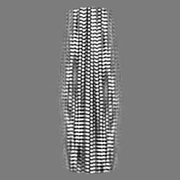
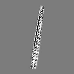
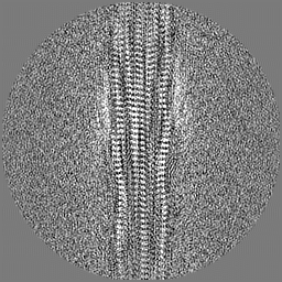
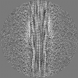
| Projections & Slices |
| ||||||||||||
| Density Histograms |


- Sample components
-Entire : Sarkosyl-extracted AppNL-G-F Abeta42 fibril
| Entire | Name: Sarkosyl-extracted AppNL-G-F Abeta42 fibril |
|---|---|
| Components |
|
-Supramolecule #1: Sarkosyl-extracted AppNL-G-F Abeta42 fibril
| Supramolecule | Name: Sarkosyl-extracted AppNL-G-F Abeta42 fibril / type: complex / ID: 1 / Parent: 0 / Macromolecule list: all Details: Fibrils purified from mouse brain labelled with Methoxy-X04 |
|---|---|
| Source (natural) | Organism: |
| Molecular weight | Theoretical: 4.441 kDa/nm |
-Macromolecule #1: Amyloid-beta precursor protein
| Macromolecule | Name: Amyloid-beta precursor protein / type: protein_or_peptide / ID: 1 Details: Humanised Abeta42 from App^NL-G-F mice with arctic mutation (E22G) Number of copies: 10 / Enantiomer: LEVO |
|---|---|
| Source (natural) | Organism: |
| Molecular weight | Theoretical: 4.448025 KDa |
| Sequence | String: DAEFRHDSGY EVHHQKLVFF AGDVGSNKGA IIGLMVGGVV IA UniProtKB: Amyloid-beta precursor protein |
-Experimental details
-Structure determination
| Method | cryo EM |
|---|---|
 Processing Processing | helical reconstruction |
| Aggregation state | filament |
-Sample preparation
| Buffer | pH: 7.4 Component:
| |||||||||
|---|---|---|---|---|---|---|---|---|---|---|
| Grid | Model: C-flat-1.2/1.3 / Material: COPPER / Mesh: 300 / Support film - Material: CARBON / Support film - topology: HOLEY / Pretreatment - Type: PLASMA CLEANING / Pretreatment - Time: 60 sec. | |||||||||
| Vitrification | Cryogen name: ETHANE / Chamber humidity: 90 % / Chamber temperature: 277 K / Instrument: FEI VITROBOT MARK IV / Details: 6s blot. | |||||||||
| Details | Sarkosyl-insoluble fibrils from App^NL-G-F mouse brain labelled with Methoxy-X04 |
- Electron microscopy
| Microscope | FEI TITAN KRIOS |
|---|---|
| Specialist optics | Energy filter - Name: TFS Selectris / Energy filter - Slit width: 10 eV |
| Image recording | Film or detector model: FEI FALCON IV (4k x 4k) / Number grids imaged: 1 / Number real images: 4165 / Average exposure time: 6.0 sec. / Average electron dose: 41.0 e/Å2 Details: 1442 raw EER frames were collected per image and combined into 40 fractions for processing |
| Electron beam | Acceleration voltage: 300 kV / Electron source:  FIELD EMISSION GUN FIELD EMISSION GUN |
| Electron optics | C2 aperture diameter: 50.0 µm / Illumination mode: FLOOD BEAM / Imaging mode: BRIGHT FIELD / Cs: 2.7 mm / Nominal defocus max: 2.9 µm / Nominal defocus min: 1.4000000000000001 µm / Nominal magnification: 130000 |
| Sample stage | Specimen holder model: FEI TITAN KRIOS AUTOGRID HOLDER |
| Experimental equipment |  Model: Titan Krios / Image courtesy: FEI Company |
-Image processing
| Final reconstruction | Applied symmetry - Helical parameters - Δz: 2.414 Å Applied symmetry - Helical parameters - Δ&Phi: 179.355 ° Applied symmetry - Helical parameters - Axial symmetry: C1 (asymmetric) Resolution.type: BY AUTHOR / Resolution: 3.2 Å / Resolution method: FSC 0.143 CUT-OFF / Software - Name: RELION (ver. 4.0) / Number images used: 3640 |
|---|---|
| Segment selection | Number selected: 136214 / Software - Name: crYOLO Details: Manually picked a subset of images to train a model for automatic fibril segment picking in crYOLO |
| Startup model | Type of model: INSILICO MODEL Details: Model generated from 2D class averages using relion_helix_inimodel2d |
| Final angle assignment | Type: NOT APPLICABLE / Software - Name: RELION (ver. 4.0) |
-Atomic model buiding 1
| Refinement | Space: REAL / Protocol: AB INITIO MODEL / Overall B value: 52 / Target criteria: Correlation coefficient |
|---|---|
| Output model | PDB-8bfb: |
