+ Open data
- Basic information
| Entry | Database: PDB / ID: 8ouo | |||||||||||||||||||||||||||||||||||||||||||||||||||||||||||||||
|---|---|---|---|---|---|---|---|---|---|---|---|---|---|---|---|---|---|---|---|---|---|---|---|---|---|---|---|---|---|---|---|---|---|---|---|---|---|---|---|---|---|---|---|---|---|---|---|---|---|---|---|---|---|---|---|---|---|---|---|---|---|---|---|---|
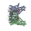
| Title | Human TPC2 in Complex with Antagonist (S)-SG-094 | |||||||||||||||||||||||||||||||||||||||||||||||||||||||||||||||
 Components Components | Two pore channel protein 2 | |||||||||||||||||||||||||||||||||||||||||||||||||||||||||||||||
 Keywords Keywords | METAL TRANSPORT / TPC2 / two-pore channel / ion channel / inhibitor / homodimer | |||||||||||||||||||||||||||||||||||||||||||||||||||||||||||||||
| Function / homology |  Function and homology information Function and homology informationendosome to lysosome transport of low-density lipoprotein particle / negative regulation of developmental pigmentation / intracellular pH reduction / intracellularly phosphatidylinositol-3,5-bisphosphate-gated monatomic cation channel activity / NAADP-sensitive calcium-release channel activity / regulation of exocytosis / melanosome membrane / endolysosome membrane / phosphatidylinositol-3,5-bisphosphate binding / response to vitamin D ...endosome to lysosome transport of low-density lipoprotein particle / negative regulation of developmental pigmentation / intracellular pH reduction / intracellularly phosphatidylinositol-3,5-bisphosphate-gated monatomic cation channel activity / NAADP-sensitive calcium-release channel activity / regulation of exocytosis / melanosome membrane / endolysosome membrane / phosphatidylinositol-3,5-bisphosphate binding / response to vitamin D / lysosome organization / ligand-gated sodium channel activity / monoatomic ion channel complex / smooth muscle contraction / voltage-gated calcium channel activity / receptor-mediated endocytosis of virus by host cell / release of sequestered calcium ion into cytosol / sodium ion transmembrane transport / calcium-mediated signaling / regulation of autophagy / Stimuli-sensing channels / calcium channel activity / intracellular calcium ion homeostasis / late endosome membrane / monoatomic ion transmembrane transport / lysosome / endosome membrane / endocytosis involved in viral entry into host cell / lysosomal membrane / protein kinase binding / identical protein binding / cytosol Similarity search - Function | |||||||||||||||||||||||||||||||||||||||||||||||||||||||||||||||
| Biological species |  Homo sapiens (human) Homo sapiens (human) | |||||||||||||||||||||||||||||||||||||||||||||||||||||||||||||||
| Method | ELECTRON MICROSCOPY / single particle reconstruction / cryo EM / Resolution: 3 Å | |||||||||||||||||||||||||||||||||||||||||||||||||||||||||||||||
 Authors Authors | Chi, G. / Pike, A.C.W. / Maclean, E.M. / Li, H. / Mukhopadhyay, S.M.M. / Bohstedt, T. / Wang, D. / McKinley, G. / Fernandez-Cid, A. / Duerr, K. | |||||||||||||||||||||||||||||||||||||||||||||||||||||||||||||||
| Funding support |  United Kingdom, 1items United Kingdom, 1items
| |||||||||||||||||||||||||||||||||||||||||||||||||||||||||||||||
 Citation Citation |  Journal: Structure / Year: 2024 Journal: Structure / Year: 2024Title: Structural basis for inhibition of the lysosomal two-pore channel TPC2 by a small molecule antagonist. Authors: Gamma Chi / Dawid Jaślan / Veronika Kudrina / Julia Böck / Huanyu Li / Ashley C W Pike / Susanne Rautenberg / Einar Krogsaeter / Tina Bohstedt / Dong Wang / Gavin McKinley / Alejandra ...Authors: Gamma Chi / Dawid Jaślan / Veronika Kudrina / Julia Böck / Huanyu Li / Ashley C W Pike / Susanne Rautenberg / Einar Krogsaeter / Tina Bohstedt / Dong Wang / Gavin McKinley / Alejandra Fernandez-Cid / Shubhashish M M Mukhopadhyay / Nicola A Burgess-Brown / Marco Keller / Franz Bracher / Christian Grimm / Katharina L Dürr /   Abstract: Two pore channels are lysosomal cation channels with crucial roles in tumor angiogenesis and viral release from endosomes. Inhibition of the two-pore channel 2 (TPC2) has emerged as potential ...Two pore channels are lysosomal cation channels with crucial roles in tumor angiogenesis and viral release from endosomes. Inhibition of the two-pore channel 2 (TPC2) has emerged as potential therapeutic strategy for the treatment of cancers and viral infections, including Ebola and COVID-19. Here, we demonstrate that antagonist SG-094, a synthetic analog of the Chinese alkaloid medicine tetrandrine with increased potency and reduced toxicity, induces asymmetrical structural changes leading to a single binding pocket at only one intersubunit interface within the asymmetrical dimer. Supported by functional characterization of mutants by Ca imaging and patch clamp experiments, we identify key residues in S1 and S4 involved in compound binding to the voltage sensing domain II. SG-094 arrests IIS4 in a downward shifted state which prevents pore opening via the IIS4/S5 linker, hence resembling gating modifiers of canonical VGICs. These findings may guide the rational development of new therapeutics antagonizing TPC2 activity. | |||||||||||||||||||||||||||||||||||||||||||||||||||||||||||||||
| History |
|
- Structure visualization
| Structure viewer | Molecule:  Molmil Molmil Jmol/JSmol Jmol/JSmol |
|---|
- Downloads & links
- Download
| PDBx/mmCIF format |  8ouo.cif.gz 8ouo.cif.gz | 280.4 KB | Display |  PDBx/mmCIF format PDBx/mmCIF format |
|---|---|---|---|---|
| PDB format |  pdb8ouo.ent.gz pdb8ouo.ent.gz | 220.5 KB | Display |  PDB format PDB format |
| PDBx/mmJSON format |  8ouo.json.gz 8ouo.json.gz | Tree view |  PDBx/mmJSON format PDBx/mmJSON format | |
| Others |  Other downloads Other downloads |
-Validation report
| Arichive directory |  https://data.pdbj.org/pub/pdb/validation_reports/ou/8ouo https://data.pdbj.org/pub/pdb/validation_reports/ou/8ouo ftp://data.pdbj.org/pub/pdb/validation_reports/ou/8ouo ftp://data.pdbj.org/pub/pdb/validation_reports/ou/8ouo | HTTPS FTP |
|---|
-Related structure data
| Related structure data |  17197MC M: map data used to model this data C: citing same article ( |
|---|---|
| Similar structure data | Similarity search - Function & homology  F&H Search F&H Search |
- Links
Links
- Assembly
| Deposited unit | 
|
|---|---|
| 1 |
|
- Components
-Protein , 1 types, 2 molecules AB
| #1: Protein | Mass: 85285.430 Da / Num. of mol.: 2 Source method: isolated from a genetically manipulated source Source: (gene. exp.)  Homo sapiens (human) / Gene: TPCN2, TPC2 / Production host: Homo sapiens (human) / Gene: TPCN2, TPC2 / Production host:  Homo sapiens (human) / References: UniProt: Q8NHX9 Homo sapiens (human) / References: UniProt: Q8NHX9 |
|---|
-Non-polymers , 5 types, 14 molecules 


| #2: Chemical | | #3: Chemical | #4: Chemical | ChemComp-W4E / ( | Mass: 451.556 Da / Num. of mol.: 1 / Source method: obtained synthetically / Formula: C30H29NO3 / Feature type: SUBJECT OF INVESTIGATION #5: Chemical | #6: Chemical | ChemComp-Y01 / |
|---|
-Details
| Has ligand of interest | Y |
|---|---|
| Has protein modification | Y |
-Experimental details
-Experiment
| Experiment | Method: ELECTRON MICROSCOPY |
|---|---|
| EM experiment | Aggregation state: PARTICLE / 3D reconstruction method: single particle reconstruction |
- Sample preparation
| Component | Name: Homodimeric complex of human Two-pore channel 2 (TPC2) Type: COMPLEX / Entity ID: #1 / Source: RECOMBINANT |
|---|---|
| Molecular weight | Value: 0.15 MDa / Experimental value: NO |
| Source (natural) | Organism:  Homo sapiens (human) Homo sapiens (human) |
| Source (recombinant) | Organism:  Homo sapiens (human) Homo sapiens (human) |
| Buffer solution | pH: 7.5 |
| Specimen | Embedding applied: NO / Shadowing applied: NO / Staining applied: NO / Vitrification applied: YES |
| Vitrification | Cryogen name: ETHANE |
- Electron microscopy imaging
| Experimental equipment |  Model: Titan Krios / Image courtesy: FEI Company |
|---|---|
| Microscopy | Model: TFS KRIOS |
| Electron gun | Electron source:  FIELD EMISSION GUN / Accelerating voltage: 300 kV / Illumination mode: FLOOD BEAM FIELD EMISSION GUN / Accelerating voltage: 300 kV / Illumination mode: FLOOD BEAM |
| Electron lens | Mode: BRIGHT FIELD / Nominal defocus max: 2400 nm / Nominal defocus min: 800 nm |
| Image recording | Electron dose: 35 e/Å2 / Film or detector model: GATAN K3 (6k x 4k) |
- Processing
| EM software | Name: PHENIX / Category: model refinement | ||||||||||||||||||||||||
|---|---|---|---|---|---|---|---|---|---|---|---|---|---|---|---|---|---|---|---|---|---|---|---|---|---|
| CTF correction | Type: PHASE FLIPPING AND AMPLITUDE CORRECTION | ||||||||||||||||||||||||
| Symmetry | Point symmetry: C1 (asymmetric) | ||||||||||||||||||||||||
| 3D reconstruction | Resolution: 3 Å / Resolution method: FSC 0.143 CUT-OFF / Num. of particles: 109417 / Symmetry type: POINT | ||||||||||||||||||||||||
| Refine LS restraints |
|
 Movie
Movie Controller


 PDBj
PDBj


