 Movie
Movie Controller
 Structure viewers
 About EMN search


-Search query
-Search result
Showing 1 - 50 of 92 items for (author: black & be)

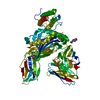
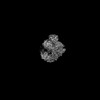
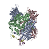
EMDB-70812: 
Tetrameric POLQ Helicase-like Domain Bound to Cmpd 19, a Small-Molecule ATPase Inhibitor and Drug Candidate Analog
Method: single particle / : Zahn KE, Scapin G

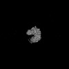
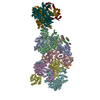
EMDB-70813:
Tetrameric POLQ Helicase-like Domain Bound to Cmpd 36, a Small-Molecule ATPase Inhibitor and Drug Candidate Analog
Method: single particle / : Zahn KE, Scapin G

PDB-9osw:
Tetrameric POLQ Helicase-like Domain Bound to Cmpd 19, a Small-Molecule ATPase Inhibitor and Drug Candidate Analog
Method: single particle / : Zahn KE, Mader P, Sicheri F

PDB-9osy:
Tetrameric POLQ Helicase-like Domain Bound to Cmpd 36, a Small-Molecule ATPase Inhibitor and Drug Candidate Analog
Method: single particle / : Zahn KE, Mader P, Sicheri F
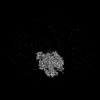
EMDB-44860:
Kinetochore on isolated human mitotic chromosome
Method: electron tomography / : Kixmoeller K, Chang YW, Black BE
EMDB-44862:
Kinetochore on isolated human mitotic chromosome after degradation of CENP-C
Method: electron tomography / : Kixmoeller K, Chang YW, Black BE

EMDB-44863:
Kinetochore on isolated human mitotic chromosome after degradation of CENP-N
Method: electron tomography / : Kixmoeller K, Chang YW, Black BE

EMDB-44864:
Kinetochore on isolated human mitotic chromosome under conditions of prolonged spindle detachment
Method: electron tomography / : Kixmoeller K, Chang YW, Black BE
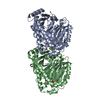
EMDB-43499:
Engineered peptide-specific binder in complex with HLA-DR1/CLIP
Method: single particle / : Jude KM, Yang X, Du H, Kassardjian A, Julien JP, Huang P, Garcia KC
PDB-8vsj:
Engineered peptide-specific binder in complex with HLA-DR1/CLIP
Method: single particle / : Jude KM, Yang X, Du H, Kassardjian A, Julien JP, Huang P, Garcia KC

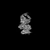
EMDB-19366:
Influenza polymerase A/H7N9-4M (ENDO(R) | Core1)
Method: single particle / : Arragain B, Cusack S

EMDB-19367:
Influenza polymerase A/H7N9-4M (ENDO(R) | Core2)
Method: single particle / : Arragain B, Cusack S

EMDB-19368:
Influenza polymerase A/H7N9-4M replication complex, an asymmetric polymerase dimer bound to human ANP32A
Method: single particle / : Arragain B, Cusack S

EMDB-19369:
Influenza polymerase A/H7N9-4M replicase minus 627(R) (from "Influenza polymerase A/H7N9-4M replication complex" | Local refinement)
Method: single particle / : Arragain B, Cusack S

EMDB-19382:
Influenza polymerase A/H7N9-4M encapsidase plus 627(R) / human ANP32A (from "Influenza polymerase A/H7N9-4M replication complex" | Local refinement)
Method: single particle / : Arragain B, Cusack S

EMDB-19383:
Influenza B polymerase, monomeric encapsidase with 5' cRNA hook bound
Method: single particle / : Arragain B, Cusack S
EMDB-19384:
Monomeric apo-influenza B polymerase, encapsidase conformation
Method: single particle / : Arragain B, Cusack S

EMDB-19385:
Pseudo-symmetrical influenza B polymerase apo-dimer, encapsidase moiety (from "Influenza B polymerase pseudo-symmetrical dimer" | Local refinement)
Method: single particle / : Arragain B, Cusack S

EMDB-19386:
Pseudo-symmetrical influenza B polymerase apo-dimer, ENDO(T) moiety (from "Influenza B polymerase pseudo-symmetrical dimer" | Local refinement)
Method: single particle / : Arragain B, Cusack S

EMDB-19387:
Pseudo-symmetrical influenza B polymerase apo-dimer, ENDO(R) moiety (from "Influenza B polymerase pseudo-symmetrical dimer" | Local refinement)
Method: single particle / : Arragain B, Cusack S
EMDB-19388:
Pseudo-symmetrical influenza B polymerase apo-dimer, ENDO(E) moiety (from "Influenza B polymerase pseudo-symmetrical dimer" | Local refinement)
Method: single particle / : Arragain B, Cusack S

EMDB-19389:
Pseudo-symmetrical influenza B polymerase apo-dimer, core-only moeity (from "Influenza B polymerase pseudo-symmetrical dimer" | Local refinement)
Method: single particle / : Arragain B, Cusack S

EMDB-19390:
Influenza B polymerase pseudo-symmetrical apo-dimer (FluPol(E)|FluPol(S))
Method: single particle / : Arragain B, Cusack S

EMDB-19391:
Influenza B polymerase, replicase (from "Influenza B polymerase apo-trimer" | Local refinement)
Method: single particle / : Arragain B, Cusack S

EMDB-19392:
Influenza B polymerase apo-trimer
Method: single particle / : Arragain B, Cusack S
EMDB-19393:
Influenza B polymerase, encapsidase plus 627(R) / human ANP32A (from "Influenza B polymerase apo-trimer" | Local refinement)
Method: single particle / : Arragain B, Cusack S

EMDB-19394:
Influenza B polymerase, replication complex, an asymmetric polymerase dimer bound to human ANP32A (from "Influenza B polymerase apo-trimer" | Local refinement)
Method: single particle / : Arragain B, Cusack S

PDB-8rmp:
Influenza polymerase A/H7N9-4M (ENDO(R) | Core1)
Method: single particle / : Arragain B, Cusack S

PDB-8rmq:
Influenza polymerase A/H7N9-4M (ENDO(R) | Core2)
Method: single particle / : Arragain B, Cusack S
PDB-8rmr:
Influenza polymerase A/H7N9-4M replication complex, an asymmetric polymerase dimer bound to human ANP32A
Method: single particle / : Arragain B, Cusack S

PDB-8rms:
Influenza polymerase A/H7N9-4M replicase minus 627(R) (from "Influenza polymerase A/H7N9-4M replication complex" | Local refinement)
Method: single particle / : Arragain B, Cusack S

PDB-8rn0:
Influenza polymerase A/H7N9-4M encapsidase plus 627(R) / human ANP32A (from "Influenza polymerase A/H7N9-4M replication complex" | Local refinement)
Method: single particle / : Arragain B, Cusack S

PDB-8rn1:
Influenza B polymerase, monomeric encapsidase with 5' cRNA hook bound
Method: single particle / : Arragain B, Cusack S

PDB-8rn2:
Monomeric apo-influenza B polymerase, encapsidase conformation
Method: single particle / : Arragain B, Cusack S
PDB-8rn3:
Pseudo-symmetrical influenza B polymerase apo-dimer, encapsidase moiety (from "Influenza B polymerase pseudo-symmetrical dimer" | Local refinement)
Method: single particle / : Arragain B, Cusack S

PDB-8rn4:
Pseudo-symmetrical influenza B polymerase apo-dimer, ENDO(T) moiety (from "Influenza B polymerase pseudo-symmetrical dimer" | Local refinement)
Method: single particle / : Arragain B, Cusack S

PDB-8rn5:
Pseudo-symmetrical influenza B polymerase apo-dimer, ENDO(R) moiety (from "Influenza B polymerase pseudo-symmetrical dimer" | Local refinement)
Method: single particle / : Arragain B, Cusack S

PDB-8rn6:
Pseudo-symmetrical influenza B polymerase apo-dimer, ENDO(E) moiety (from "Influenza B polymerase pseudo-symmetrical dimer" | Local refinement)
Method: single particle / : Arragain B, Cusack S

PDB-8rn7:
Pseudo-symmetrical influenza B polymerase apo-dimer, core-only moeity (from "Influenza B polymerase pseudo-symmetrical dimer" | Local refinement)
Method: single particle / : Arragain B, Cusack S

PDB-8rn8:
Influenza B polymerase pseudo-symmetrical apo-dimer (FluPol(E)|FluPol(S))
Method: single particle / : Arragain B, Cusack S

PDB-8rn9:
Influenza B polymerase, replicase (from "Influenza B polymerase apo-trimer" | Local refinement)
Method: single particle / : Arragain B, Cusack S
PDB-8rna:
Influenza B polymerase apo-trimer
Method: single particle / : Arragain B, Cusack S

PDB-8rnb:
Influenza B polymerase, encapsidase plus 627(R) / human ANP32A (from "Influenza B polymerase apo-trimer" | Local refinement)
Method: single particle / : Arragain B, Cusack S

PDB-8rnc:
Influenza B polymerase, replication complex, an asymmetric polymerase dimer bound to human ANP32A (from "Influenza B polymerase apo-trimer" | Local refinement)
Method: single particle / : Arragain B, Cusack S

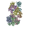
EMDB-40480:
TUBB4B and TUBA1A Heterodimer from Human Respiratory Doublet Microtubules
Method: single particle / : Anderson JR, Gui M, Brown A
PDB-8sh7:
TUBB4B and TUBA1A Heterodimer from Human Respiratory Doublet Microtubules
Method: single particle / : Anderson JR, Gui M, Brown A

EMDB-43330:
Tomogram of human Alzheimer's disease brain tissue showing variety of membrane vesicles and axonal cross-sections
Method: electron tomography / : Creekmore BC, Lee EB, Chang YW

EMDB-43331:
Tomogram of human Alzheimer's disease brain tissue showing longitudinal axonal cross-section
Method: electron tomography / : Creekmore BC, Lee EB, Chang YW

EMDB-43334:
Tomogram of human Alzheimer's disease brain tissue showing variety of membrane vesicles
Method: electron tomography / : Creekmore BC, Lee EB, Chang YW
EMDB-16847:
CryoEM structure of holo e4D2
Method: single particle / : Yadav KNS, Hutchins G, Berger Schaffitzel C, Anderson R
Pages:
 wwPDB to switch to version 3 of the EMDB data model
