 Movie
Movie Controller

[English] 日本語
 Yorodumi
- EMDB-3610: Malaria-infected red blood cell section showing schizont stalled ... -
+ Open data

- Basic information
| Entry | Database: EMDB / ID: EMD-3610 | |||||||||
|---|---|---|---|---|---|---|---|---|---|---|
| Title | Malaria-infected red blood cell section showing schizont stalled in egress with the inhibitor E64 | |||||||||
 Map data Map data | Resin section tomogram of a Plasmodium falciparum infected erythrocyte treated with the egress inhibitor E64 | |||||||||
 Sample Sample |
| |||||||||
| Biological species |  | |||||||||
| Method | electron tomography / negative staining | |||||||||
 Authors Authors | Hale VL / Saibil HR | |||||||||
 Citation Citation | Journal: Proc Natl Acad Sci U S A / Year: 2017 Title: Parasitophorous vacuole poration precedes its rupture and rapid host erythrocyte cytoskeleton collapse in egress. Authors: Victoria L Hale / Jean M Watermeyer / Fiona Hackett / Gema Vizcay-Barrena / Christiaan van Ooij / James A Thomas / Matthew C Spink / Maria Harkiolaki / Elizabeth Duke / Roland A Fleck / ...Authors: Victoria L Hale / Jean M Watermeyer / Fiona Hackett / Gema Vizcay-Barrena / Christiaan van Ooij / James A Thomas / Matthew C Spink / Maria Harkiolaki / Elizabeth Duke / Roland A Fleck / Michael J Blackman / Helen R Saibil /  Abstract: In the asexual blood stages of malarial infection, merozoites invade erythrocytes and replicate within a parasitophorous vacuole to form daughter cells that eventually exit (egress) by sequential ...In the asexual blood stages of malarial infection, merozoites invade erythrocytes and replicate within a parasitophorous vacuole to form daughter cells that eventually exit (egress) by sequential rupture of the vacuole and erythrocyte membranes. The current model is that PKG, a malarial cGMP-dependent protein kinase, triggers egress, activating malarial proteases and other effectors. Using selective inhibitors of either PKG or cysteine proteases to separately inhibit the sequential steps in membrane perforation, combined with video microscopy, electron tomography, electron energy loss spectroscopy, and soft X-ray tomography of mature intracellular parasites, we resolve intermediate steps in egress. We show that the parasitophorous vacuole membrane (PVM) is permeabilized 10-30 min before its PKG-triggered breakdown into multilayered vesicles. Just before PVM breakdown, the host red cell undergoes an abrupt, dramatic shape change due to the sudden breakdown of the erythrocyte cytoskeleton, before permeabilization and eventual rupture of the erythrocyte membrane to release the parasites. In contrast to the previous view of PKG-triggered initiation of egress and a gradual dismantling of the host erythrocyte cytoskeleton over the course of schizont development, our findings identify an initial step in egress and show that host cell cytoskeleton breakdown is restricted to a narrow time window within the final stages of egress. | |||||||||
| History |
|
- Structure visualization
| Movie |
 Movie viewer Movie viewer |
|---|---|
| Structure viewer | EM map: SurfViewMolmilJmol/JSmol |
| Supplemental images |

- Downloads & links
-EMDB archive
| Map data | emd_3610.map.gz | 536.4 MB | EMDB map data format | |
|---|---|---|---|---|
| Header (meta data) | emd-3610-v30.xmlemd-3610.xml | 10.2 KB 10.2 KB | Display Display | EMDB header |
| Images |  emd_3610.png emd_3610.png | 292.5 KB | ||
| Archive directory |  http://ftp.pdbj.org/pub/emdb/structures/EMD-3610ftp://ftp.pdbj.org/pub/emdb/structures/EMD-3610 http://ftp.pdbj.org/pub/emdb/structures/EMD-3610ftp://ftp.pdbj.org/pub/emdb/structures/EMD-3610 | HTTPS FTP |
-Related structure data
| Related structure data | 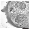 3586C 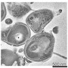 3587C 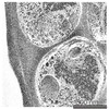 3606C C: citing same article ( |
|---|---|
| EM raw data | EMPIAR-10087 (Title: Soft X-ray tomography of Plasmodium falciparum infected human erythrocytes stalled in egress by the inhibitors Compound 2 and E64 Data size: 280.6 MB Data #1: Soft X-ray tomograms of Plasmodium falciparum infected human erythrocytes stalled in egress [Soft X-ray tomograms]) |
-Links
| EMDB pages | EMDB (EBI/PDBe) / EMDataResource |
|---|
-Map
| File | Download / File: emd_3610.map.gz / Format: CCP4 / Size: 877.9 MB / Type: IMAGE STORED AS SIGNED BYTE | ||||||||||||||||||||||||||||||||||||||||||||||||||||||||||||
|---|---|---|---|---|---|---|---|---|---|---|---|---|---|---|---|---|---|---|---|---|---|---|---|---|---|---|---|---|---|---|---|---|---|---|---|---|---|---|---|---|---|---|---|---|---|---|---|---|---|---|---|---|---|---|---|---|---|---|---|---|---|
| Annotation | Resin section tomogram of a Plasmodium falciparum infected erythrocyte treated with the egress inhibitor E64 | ||||||||||||||||||||||||||||||||||||||||||||||||||||||||||||
| Voxel size | X=Y=Z: 9.30825 Å | ||||||||||||||||||||||||||||||||||||||||||||||||||||||||||||
| Density |
| ||||||||||||||||||||||||||||||||||||||||||||||||||||||||||||
| Symmetry | Space group: 1 | ||||||||||||||||||||||||||||||||||||||||||||||||||||||||||||
| Details | EMDB XML:
CCP4 map header:
| ||||||||||||||||||||||||||||||||||||||||||||||||||||||||||||
-Supplemental data
- Sample components
-Entire : Plasmodium falciparum infected human erythrocyte treated with the...
| Entire | Name: Plasmodium falciparum infected human erythrocyte treated with the egress inhibitor E64 |
|---|---|
| Components |
|
-Supramolecule #1: Plasmodium falciparum infected human erythrocyte treated with the...
| Supramolecule | Name: Plasmodium falciparum infected human erythrocyte treated with the egress inhibitor E64 type: cell / ID: 1 / Parent: 0 Details: High pressure frozen and freeze-substituted cell section |
|---|---|
| Source (natural) | Organism: |
-Experimental details
-Structure determination
| Method | negative staining |
|---|---|
 Processing Processing | electron tomography |
| Aggregation state | cell |
-Sample preparation
| Buffer | pH: 7 / Details: RPMI medium |
|---|---|
| Staining | Type: NEGATIVE / Material: Uranyl Acetate |
| Sugar embedding | Material: HM20 / Details: Freeze substitution |
| Grid | Material: COPPER / Support film - Material: CARBON / Support film - topology: CONTINUOUS / Pretreatment - Type: GLOW DISCHARGE |
| High pressure freezing | Instrument: OTHER Details: The value given for _emd_high_pressure_freezing.instrument is Baltech HPM010. This is not in a list of allowed values set(['LEICA EM PACT2', 'LEICA EM PACT', 'EMS-002 RAPID IMMERSION ...Details: The value given for _emd_high_pressure_freezing.instrument is Baltech HPM010. This is not in a list of allowed values set(['LEICA EM PACT2', 'LEICA EM PACT', 'EMS-002 RAPID IMMERSION FREEZER', 'OTHER', 'LEICA EM HPM100', 'BAL-TEC HPM 010']) so OTHER is written into the XML file. |
| Sectioning | Ultramicrotomy - Instrument: Leica EM UC7 / Ultramicrotomy - Temperature: 293 K / Ultramicrotomy - Final thickness: 250 |
| Fiducial marker | Manufacturer: Electron microscopy sciences / Diameter: 10 nm |
- Electron microscopy
| Microscope | FEI TECNAI F20 |
|---|---|
| Image recording | Film or detector model: GATAN ULTRASCAN 4000 (4k x 4k) / Average electron dose: 100.0 e/Å2 |
| Electron beam | Acceleration voltage: 200 kV / Electron source:  FIELD EMISSION GUN FIELD EMISSION GUN |
| Electron optics | Illumination mode: FLOOD BEAM / Imaging mode: BRIGHT FIELD |
| Sample stage | Specimen holder model: OTHER |
| Experimental equipment |  Model: Tecnai F20 / Image courtesy: FEI Company |
-Image processing
| Final reconstruction | Algorithm: BACK PROJECTION / Software - Name: IMOD / Number images used: 131 |
|---|
