 Movie
Movie Controller
 Structure viewers
 About Yorodumi Papers


+Search query
-Structure paper
| Title | Parasitophorous vacuole poration precedes its rupture and rapid host erythrocyte cytoskeleton collapse in egress. |
|---|---|
| Journal, issue, pages | Proc Natl Acad Sci U S A, Vol. 114, Issue 13, Page 3439-3444, Year 2017 |
| Publish date | Mar 28, 2017 |
 Authors Authors | Victoria L Hale / Jean M Watermeyer / Fiona Hackett / Gema Vizcay-Barrena / Christiaan van Ooij / James A Thomas / Matthew C Spink / Maria Harkiolaki / Elizabeth Duke / Roland A Fleck / Michael J Blackman / Helen R Saibil /  |
| PubMed Abstract | In the asexual blood stages of malarial infection, merozoites invade erythrocytes and replicate within a parasitophorous vacuole to form daughter cells that eventually exit (egress) by sequential ...In the asexual blood stages of malarial infection, merozoites invade erythrocytes and replicate within a parasitophorous vacuole to form daughter cells that eventually exit (egress) by sequential rupture of the vacuole and erythrocyte membranes. The current model is that PKG, a malarial cGMP-dependent protein kinase, triggers egress, activating malarial proteases and other effectors. Using selective inhibitors of either PKG or cysteine proteases to separately inhibit the sequential steps in membrane perforation, combined with video microscopy, electron tomography, electron energy loss spectroscopy, and soft X-ray tomography of mature intracellular parasites, we resolve intermediate steps in egress. We show that the parasitophorous vacuole membrane (PVM) is permeabilized 10-30 min before its PKG-triggered breakdown into multilayered vesicles. Just before PVM breakdown, the host red cell undergoes an abrupt, dramatic shape change due to the sudden breakdown of the erythrocyte cytoskeleton, before permeabilization and eventual rupture of the erythrocyte membrane to release the parasites. In contrast to the previous view of PKG-triggered initiation of egress and a gradual dismantling of the host erythrocyte cytoskeleton over the course of schizont development, our findings identify an initial step in egress and show that host cell cytoskeleton breakdown is restricted to a narrow time window within the final stages of egress. |
 External links External links | Proc Natl Acad Sci U S A / PubMed:28292906 / PubMed Central |
| Methods | EM (tomography) |
| Structure data | 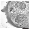 EMDB-3586: 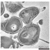 EMDB-3587: 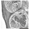 EMDB-3606: 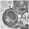 EMDB-3610: |
| Source |
|

