Natural Sciences and Engineering Research Council (Canada)
RGPIN-2016-04954
Canada
Natural Sciences and Engineering Research Council (Canada)
RGPIN-2018-04813
Canada
Canadian Institutes of Health Research
PJT-156354
Canada
Citation
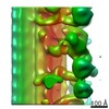
Journal: Elife / Year: 2020 Title: The inner junction complex of the cilia is an interaction hub that involves tubulin post-translational modifications. Authors: Ahmad Abdelzaher Zaki Khalifa / Muneyoshi Ichikawa / Daniel Dai / Shintaroh Kubo / Corbin Steven Black / Katya Peri / Thomas S McAlear / Simon Veyron / Shun Kai Yang / Javier Vargas / ...Authors: Ahmad Abdelzaher Zaki Khalifa / Muneyoshi Ichikawa / Daniel Dai / Shintaroh Kubo / Corbin Steven Black / Katya Peri / Thomas S McAlear / Simon Veyron / Shun Kai Yang / Javier Vargas / Susanne Bechstedt / Jean-François Trempe / Khanh Huy Bui / Abstract: Microtubules are cytoskeletal structures involved in stability, transport and organization in the cell. The building blocks, the α- and β-tubulin heterodimers, form protofilaments that associate ...Microtubules are cytoskeletal structures involved in stability, transport and organization in the cell. The building blocks, the α- and β-tubulin heterodimers, form protofilaments that associate laterally into the hollow microtubule. Microtubule also exists as highly stable doublet microtubules in the cilia where stability is needed for ciliary beating and function. The doublet microtubule maintains its stability through interactions at its inner and outer junctions where its A- and B-tubules meet. Here, using cryo-electron microscopy, bioinformatics and mass spectrometry of the doublets of and , we identified two new inner junction proteins, FAP276 and FAP106, and an inner junction-associated protein, FAP126, thus presenting the complete answer to the inner junction identity and localization. Our structural study of the doublets shows that the inner junction serves as an interaction hub that involves tubulin post-translational modifications. These interactions contribute to the stability of the doublet and hence, normal ciliary motility.
History
Deposition
Oct 21, 2019
-
Header (metadata) release
Oct 30, 2019
-
Map release
Jan 29, 2020
-
Update
Jan 29, 2020
-
Current status
Jan 29, 2020
Processing site: RCSB / Status: Released
-
Structure visualization
Movie
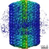
Surface view with section colored by density value
Model: Quantifoil R2/2 / Material: COPPER / Mesh: 200 / Support film - Material: CARBON / Support film - topology: HOLEY / Pretreatment - Type: GLOW DISCHARGE
Vitrification
Cryogen name: ETHANE / Chamber humidity: 100 % / Chamber temperature: 298 K / Instrument: FEI VITROBOT MARK IV / Details: Blot force 3 for 5 seconds.
-
Electron microscopy
Microscope
FEI TITAN KRIOS
Image recording
Film or detector model: FEI FALCON II (4k x 4k) / Detector mode: INTEGRATING / Digitization - Frames/image: 1-7 / Number real images: 9528 / Average exposure time: 1.4 sec. / Average electron dose: 45.0 e/Å2
Electron beam
Acceleration voltage: 300 kV / Electron source: FIELD EMISSION GUN
In the structure databanks used in Yorodumi, some data are registered as the other names, "COVID-19 virus" and "2019-nCoV". Here are the details of the virus and the list of structure data.
Jan 31, 2019. EMDB accession codes are about to change! (news from PDBe EMDB page)
EMDB accession codes are about to change! (news from PDBe EMDB page)
The allocation of 4 digits for EMDB accession codes will soon come to an end. Whilst these codes will remain in use, new EMDB accession codes will include an additional digit and will expand incrementally as the available range of codes is exhausted. The current 4-digit format prefixed with “EMD-” (i.e. EMD-XXXX) will advance to a 5-digit format (i.e. EMD-XXXXX), and so on. It is currently estimated that the 4-digit codes will be depleted around Spring 2019, at which point the 5-digit format will come into force.
The EM Navigator/Yorodumi systems omit the EMD- prefix.
Related info.:Q: What is EMD? / ID/Accession-code notation in Yorodumi/EM Navigator
Yorodumi is a browser for structure data from EMDB, PDB, SASBDB, etc.
This page is also the successor to EM Navigator detail page, and also detail information page/front-end page for Omokage search.
The word "yorodu" (or yorozu) is an old Japanese word meaning "ten thousand". "mi" (miru) is to see.
Related info.:EMDB / PDB / SASBDB / Comparison of 3 databanks / Yorodumi Search / Aug 31, 2016. New EM Navigator & Yorodumi / Yorodumi Papers / Jmol/JSmol / Function and homology information / Changes in new EM Navigator and Yorodumi
 Movie
Movie Controller

 Yorodumi
 Open data
 Basic information
 Map data
 Sample
 Function and homology information
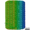
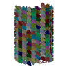
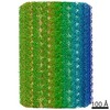
 Chlamydomonas reinhardtii (plant)
 Authors
 Canada, 3 items
  Citation
 Structure visualization
 Downloads & links
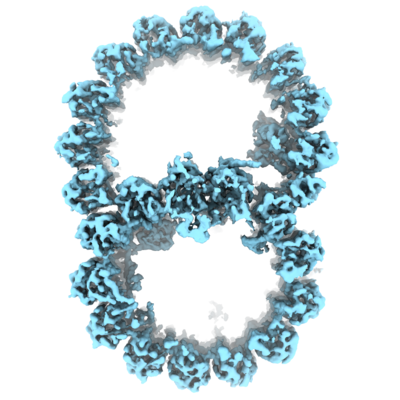 emd_20855.png
 http://ftp.pdbj.org/pub/emdb/structures/EMD-20855


 Z (Sec.)
 Y (Row.)
 X (Col.)


 Sample components
 Processing
 Electron microscopy
 FIELD EMISSION GUN

