 Movie
Movie Controller


+ Open data

- Basic information
| Entry | Database: PDB / ID: 6u42 | ||||||
|---|---|---|---|---|---|---|---|
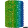
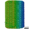
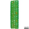
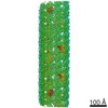
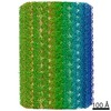
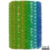
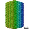
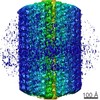
| Title | Natively decorated ciliary doublet microtubule | ||||||
 Components Components |
| ||||||
 Keywords Keywords | PROTEIN FIBRIL / motile cilia / microtubule doublet / repetitive structure | ||||||
| Function / homology |  Function and homology information Function and homology informationaxonemal central pair / axonemal doublet microtubule / positive regulation of cilium-dependent cell motility / organelle / outer dynein arm / outer dynein arm assembly / cilium-dependent cell motility / regulation of cilium beat frequency involved in ciliary motility / establishment of protein localization to organelle / cilium movement involved in cell motility ...axonemal central pair / axonemal doublet microtubule / positive regulation of cilium-dependent cell motility / organelle / outer dynein arm / outer dynein arm assembly / cilium-dependent cell motility / regulation of cilium beat frequency involved in ciliary motility / establishment of protein localization to organelle / cilium movement involved in cell motility / cilium movement / axoneme assembly / axonemal microtubule / negative regulation of microtubule depolymerization / nucleoside-diphosphate kinase / UTP biosynthetic process / CTP biosynthetic process / motile cilium / nucleoside diphosphate kinase activity / microtubule associated complex / GTP biosynthetic process / mitotic cytokinesis / cilium assembly / axoneme / alpha-tubulin binding / microtubule-based process / Hsp70 protein binding / mitotic spindle organization / ATP-dependent protein folding chaperone / meiotic cell cycle / Hsp90 protein binding / structural constituent of cytoskeleton / mitotic spindle / microtubule / Hydrolases; Acting on acid anhydrides; Acting on GTP to facilitate cellular and subcellular movement / cytoskeleton / calmodulin binding / cilium / ciliary basal body / hydrolase activity / GTPase activity / calcium ion binding / GTP binding / ATP binding / metal ion binding / nucleus Similarity search - Function | ||||||
| Biological species |   Chlamydomonas reinhardtii (plant) Chlamydomonas reinhardtii (plant) | ||||||
| Method | ELECTRON MICROSCOPY / single particle reconstruction / cryo EM / Resolution: 3.4 Å | ||||||
 Authors Authors | Ma, M. / Stoyanova, M. / Rademacher, G. / Dutcher, S.K. / Brown, A. / Zhang, R. | ||||||
| Funding support |  United States, 1items United States, 1items
| ||||||
 Citation Citation | Journal: Cell / Year: 2019 Title: Structure of the Decorated Ciliary Doublet Microtubule. Authors: Meisheng Ma / Mihaela Stoyanova / Griffin Rademacher / Susan K Dutcher / Alan Brown / Rui Zhang / Abstract: The axoneme of motile cilia is the largest macromolecular machine of eukaryotic cells. In humans, impaired axoneme function causes a range of ciliopathies. Axoneme assembly, structure, and motility ...The axoneme of motile cilia is the largest macromolecular machine of eukaryotic cells. In humans, impaired axoneme function causes a range of ciliopathies. Axoneme assembly, structure, and motility require a radially arranged set of doublet microtubules, each decorated in repeating patterns with non-tubulin components. We use single-particle cryo-electron microscopy to visualize and build an atomic model of the repeating structure of a native axonemal doublet microtubule, which reveals the identities, positions, repeat lengths, and interactions of 38 associated proteins, including 33 microtubule inner proteins (MIPs). The structure demonstrates how these proteins establish the unique architecture of doublet microtubules, maintain coherent periodicities along the axoneme, and stabilize the microtubules against the repeated mechanical stress induced by ciliary motility. Our work elucidates the architectural principles that underpin the assembly of this large, repetitive eukaryotic structure and provides a molecular basis for understanding the etiology of human ciliopathies. | ||||||
| History |
|
- Structure visualization
| Movie |
Movie viewer |
|---|---|
| Structure viewer | Molecule: MolmilJmol/JSmol |
- Downloads & links
-Download
| PDBx/mmCIF format | 6u42.cif.gz | 28.3 MB | Display | PDBx/mmCIF format |
|---|---|---|---|---|
| PDB format | pdb6u42.ent.gz | Display | PDB format | |
| PDBx/mmJSON format | 6u42.json.gz | Tree view | PDBx/mmJSON format | |
| Others |  Other downloads Other downloads |
-Validation report
| Arichive directory | https://data.pdbj.org/pub/pdb/validation_reports/u4/6u42ftp://data.pdbj.org/pub/pdb/validation_reports/u4/6u42 | HTTPS FTP |
|---|
-Related structure data
| Related structure data | 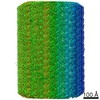 20631MC M: map data used to model this data C: citing same article ( |
|---|---|
| Similar structure data |

-Links
 PDBj
PDBj


- Assembly
| Deposited unit | 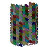
|
|---|---|
| 1 |
|
-Components
+Protein , 38 types, 451 molecules 13579A1A3A5A7A9B1B3B5B8C0C2C4C6C8D0D2D4D6D8E0E2E4E6E8F0...
-Non-polymers , 3 types, 510 molecules 


| #39: Chemical | ChemComp-GDP /  Type: RNA linking / Mass: 443.201 Da / Num. of mol.: 170 / Source method: obtained synthetically / Formula: C10H15N5O11P2 / Comment: GDP, energy-carrying molecule*YM Type: RNA linking / Mass: 443.201 Da / Num. of mol.: 170 / Source method: obtained synthetically / Formula: C10H15N5O11P2 / Comment: GDP, energy-carrying molecule*YM#40: Chemical | ChemComp-MG /  Mass: 24.305 Da / Num. of mol.: 170 / Source method: obtained synthetically / Formula: Mg Mass: 24.305 Da / Num. of mol.: 170 / Source method: obtained synthetically / Formula: Mg#41: Chemical | ChemComp-GTP /  Mass: 523.180 Da / Num. of mol.: 170 / Source method: obtained synthetically / Formula: C10H16N5O14P3 / Comment: GTP, energy-carrying molecule*YM Mass: 523.180 Da / Num. of mol.: 170 / Source method: obtained synthetically / Formula: C10H16N5O14P3 / Comment: GTP, energy-carrying molecule*YM |
|---|
-Details
| Compound details | The authors state that there are eight copies of the RIB72 protein, each connected by a flexible ...The authors state that there are eight copies of the RIB72 protein, each connected by a flexible linker to an N-terminus. They are not sure which N-terminus belongs to which protein. The N-terminus that has been assigned to chain 6U could, therefore, belong to another molecule or even to one not in their reconstruction. |
|---|---|
| Has ligand of interest | N |
| Has protein modification | Y |
-Experimental details
-Experiment
| Experiment | Method: ELECTRON MICROSCOPY |
|---|---|
| EM experiment | Aggregation state: FILAMENT / 3D reconstruction method: single particle reconstruction |
- Sample preparation
| Component | Name: Doublet microtubule from wild-type Chlamydomonas reinhardtii Type: COMPLEX / Entity ID: #1-#38 / Source: NATURAL |
|---|---|
| Molecular weight | Experimental value: NO |
| Source (natural) | Organism: Chlamydomonas reinhardtii (plant) / Strain: CC-125 |
| Buffer solution | pH: 7.4 Details: 30 mM HEPES, 5 mM MgSO4, 1 mM DTT, 0.5 mM EGTA, 25 mM KCl, PH 7.4 |
| Buffer component | Name: HMDEKP |
| Specimen | Embedding applied: NO / Shadowing applied: NO / Staining applied: NO / Vitrification applied: YES |
| Vitrification | Instrument: FEI VITROBOT MARK IV / Cryogen name: ETHANE / Humidity: 95 % / Chamber temperature: 277 K / Details: blot for 4 seconds before plunging |
- Electron microscopy imaging
| Experimental equipment |  Model: Titan Krios / Image courtesy: FEI Company |
|---|---|
| Microscopy | Model: FEI TITAN KRIOS |
| Electron gun | Electron source:  FIELD EMISSION GUN / Accelerating voltage: 300 kV / Illumination mode: FLOOD BEAM FIELD EMISSION GUN / Accelerating voltage: 300 kV / Illumination mode: FLOOD BEAM |
| Electron lens | Mode: BRIGHT FIELD / Nominal magnification: 81000 X / Calibrated magnification: 81000 X / Nominal defocus max: 2750 nm / Nominal defocus min: 1250 nm / Calibrated defocus min: 1000 nm / Calibrated defocus max: 3100 nm / Cs: 0.01 mm / Alignment procedure: ZEMLIN TABLEAU |
| Specimen holder | Cryogen: NITROGEN / Specimen holder model: FEI TITAN KRIOS AUTOGRID HOLDER |
| Image recording | Average exposure time: 9 sec. / Electron dose: 38.9 e/Å2 / Detector mode: COUNTING / Film or detector model: GATAN K2 QUANTUM (4k x 4k) / Num. of grids imaged: 6 / Num. of real images: 8314 |
| EM imaging optics | Energyfilter name: GIF Bioquantum / Energyfilter slit width: 20 eV Spherical aberration corrector: Microscope is equipped with a Cs corrector |
| Image scans | Width: 3838 / Height: 3710 / Movie frames/image: 30 / Used frames/image: 1-30 |
- Processing
| EM software |
| ||||||||||||||||||||||||||||||
|---|---|---|---|---|---|---|---|---|---|---|---|---|---|---|---|---|---|---|---|---|---|---|---|---|---|---|---|---|---|---|---|
| Image processing | Details: The movies were drift-corrected using UCSF MotionCorr2 | ||||||||||||||||||||||||||||||
| CTF correction | Type: PHASE FLIPPING AND AMPLITUDE CORRECTION | ||||||||||||||||||||||||||||||
| Particle selection | Details: doublet microtubules were manually selected | ||||||||||||||||||||||||||||||
| Symmetry | Point symmetry: C1 (asymmetric) | ||||||||||||||||||||||||||||||
| 3D reconstruction | Resolution: 3.4 Å / Resolution method: FSC 0.143 CUT-OFF / Num. of particles: 143765 / Symmetry type: POINT | ||||||||||||||||||||||||||||||
| Atomic model building | B value: 50 / Protocol: AB INITIO MODEL / Space: REAL |
