axonemal central pair / axonemal doublet microtubule / positive regulation of cilium-dependent cell motility / organelle / outer dynein arm / outer dynein arm assembly / cilium-dependent cell motility / regulation of cilium beat frequency involved in ciliary motility / establishment of protein localization to organelle / cilium movement involved in cell motility ...axonemal central pair / axonemal doublet microtubule / positive regulation of cilium-dependent cell motility / organelle / outer dynein arm / outer dynein arm assembly / cilium-dependent cell motility / regulation of cilium beat frequency involved in ciliary motility / establishment of protein localization to organelle / cilium movement involved in cell motility / axoneme assembly / cilium movement / axonemal microtubule / negative regulation of microtubule depolymerization / nucleoside-diphosphate kinase / UTP biosynthetic process / CTP biosynthetic process / motile cilium / nucleoside diphosphate kinase activity / microtubule associated complex / GTP biosynthetic process / mitotic cytokinesis / cilium assembly / axoneme / alpha-tubulin binding / microtubule-based process / Hsp70 protein binding / mitotic spindle organization / meiotic cell cycle / ATP-dependent protein folding chaperone / Hsp90 protein binding / structural constituent of cytoskeleton / mitotic spindle / cytoskeleton / microtubule / Hydrolases; Acting on acid anhydrides; Acting on GTP to facilitate cellular and subcellular movement / calmodulin binding / cilium / ciliary basal body / hydrolase activity / GTPase activity / calcium ion binding / GTP binding / ATP binding / metal ion binding / nucleus Similarity search - Function
: / WDR90, 4th beta-propeller / : / : / ODAD1 central coiled coil region / Protein Flattop / Flattop / Nucleoside diphosphate kinase 7 / Piercer of microtubule wall 1/2 / Piercer of microtubule wall 1/2 ...: / WDR90, 4th beta-propeller / : / : / ODAD1 central coiled coil region / Protein Flattop / Flattop / Nucleoside diphosphate kinase 7 / Piercer of microtubule wall 1/2 / Piercer of microtubule wall 1/2 / Cilia- and flagella-associated protein 141 / Cilia- and flagella-associated protein 141 / Cilia- and flagella-associated protein 161-like domain / : / : / : / Nucleoside diphosphate kinase 7 N-terminal PH domain / Meiosis-specific nuclear structural protein 1 / Cilia- and flagella-associated protein 45 / NDPK7, second NDPk domain / Cilia- and flagella- associated protein 210 / CFAP53/TCHP / Trichohyalin-plectin-homology domain / Trichohyalin-plectin-homology domain / RIB43A / RIB43A / DM10 domain / EF-hand domain-containing protein EFHC1/EFHC2/EFHB / DM10 domain / DM10 domain profile. / Domains in hypothetical proteins in Drosophila, C. elegans and mammals. Occurs singly in some nucleoside diphosphate kinases. / Enkurin domain / : / Calmodulin-binding / Enkurin domain profile. / : / CFA20 domain / Cilia- and flagella-associated protein 20/CFAP20DC / CFA20 domain / : / Parkin co-regulated protein / Parkin co-regulated protein / EF-hand domain / Nucleoside diphosphate kinase, active site / Nucleoside diphosphate kinase (NDPK) active site signature. / Nucleoside diphosphate kinase / Nucleoside diphosphate kinase (NDPK)-like domain profile. / Nucleoside diphosphate kinase-like domain / Nucleoside diphosphate kinase / NDK / Nucleoside diphosphate kinase-like domain superfamily / Heat shock protein 70kD, peptide-binding domain superfamily / Heat shock protein 70 family / Hsp70 protein / Alpha tubulin / Tubulin-beta mRNA autoregulation signal. / Beta tubulin, autoregulation binding site / Beta tubulin / Tubulin / Tubulin, C-terminal / Tubulin C-terminal domain / Tubulin, conserved site / Tubulin subunits alpha, beta, and gamma signature. / Nucleotide-diphospho-sugar transferases / Tubulin/FtsZ family, C-terminal domain / Tubulin/FtsZ-like, C-terminal domain / Tubulin/FtsZ, C-terminal / Tubulin/FtsZ, 2-layer sandwich domain / Tubulin/FtsZ family, GTPase domain / Tubulin/FtsZ family, GTPase domain / Tubulin/FtsZ, GTPase domain / Tubulin/FtsZ, GTPase domain superfamily / EF-hand domain pair / EF-hand, calcium binding motif / EF-Hand 1, calcium-binding site / EF-hand calcium-binding domain. / EF-hand calcium-binding domain profile. / EF-hand domain / EF-hand domain pair / WD domain, G-beta repeat / Armadillo-type fold / WD40 repeat, conserved site / Trp-Asp (WD) repeats signature. / Trp-Asp (WD) repeats profile. / Trp-Asp (WD) repeats circular profile. / WD40 repeats / WD40 repeat / WD40-repeat-containing domain superfamily / WD40/YVTN repeat-like-containing domain superfamily Similarity search - Domain/homology
Meiosis-specific nuclear structural protein 1 / Cilia- and flagella-associated protein 52 / Uncharacterized protein / Uncharacterized protein / Uncharacterized protein / Flagellar associated protein / Trichohyalin-plectin-homology domain-containing protein / Uncharacterized protein / Uncharacterized protein / Uncharacterized protein ...Meiosis-specific nuclear structural protein 1 / Cilia- and flagella-associated protein 52 / Uncharacterized protein / Uncharacterized protein / Uncharacterized protein / Flagellar associated protein / Trichohyalin-plectin-homology domain-containing protein / Uncharacterized protein / Uncharacterized protein / Uncharacterized protein / Uncharacterized protein / Uncharacterized protein / EF-hand domain-containing protein / Cilia- and flagella-associated protein 45 / Uncharacterized protein / Flagellar associated protein / Uncharacterized protein / Cilia- and flagella-associated protein 53 / Cilia- and flagella-associated protein 20 / Protein Flattop homolog / Uncharacterized protein / Uncharacterized protein / Uncharacterized protein / Uncharacterized protein / Uncharacterized protein / Uncharacterized protein / Uncharacterized protein / Uncharacterized protein / Outer dynein arm protein 1 / Nucleoside diphosphate kinase / Uncharacterized protein / Parkin-co-regulated gene product / Tubulin beta-1/beta-2 chain / Tubulin alpha-1 chain / Outer dynein arm-docking complex protein DC3 / RIB72 protein / Flagellar protofilament ribbon protein Similarity search - Component
Biological species
Chlamydomonas reinhardtii (plant)
Method
single particle reconstruction / cryo EM / Resolution: 3.4 Å
National Institutes of Health/National Institute of General Medical Sciences (NIH/NIGMS)
R01GM032843
United States
Citation
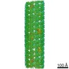
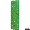
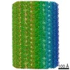
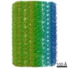
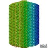
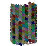
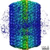
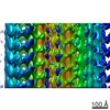
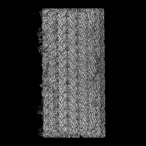
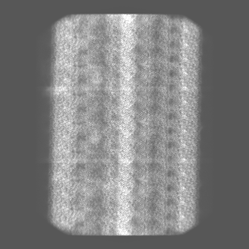
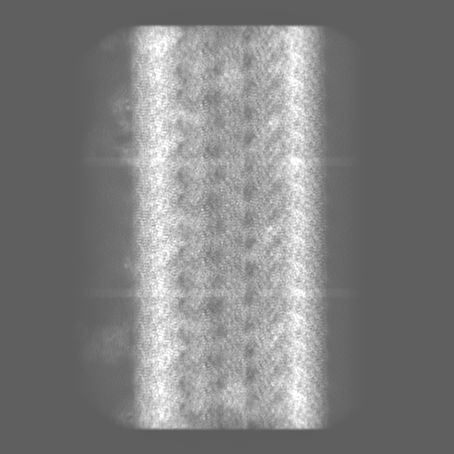
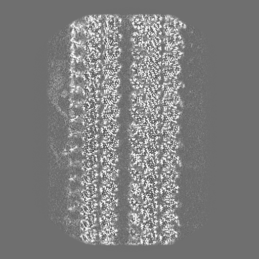
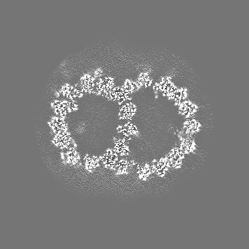
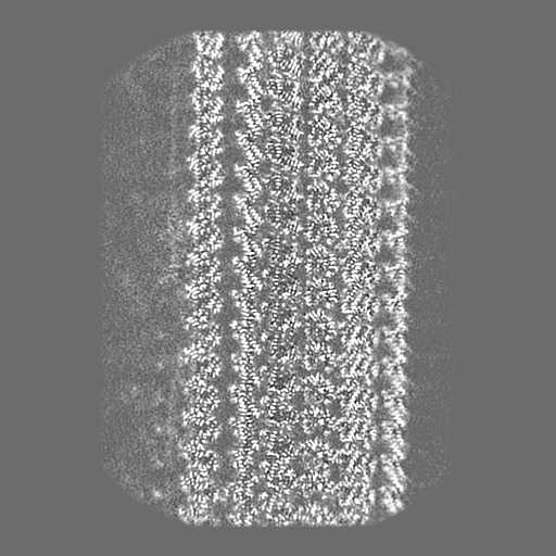
Journal: Cell / Year: 2019 Title: Structure of the Decorated Ciliary Doublet Microtubule. Authors: Meisheng Ma / Mihaela Stoyanova / Griffin Rademacher / Susan K Dutcher / Alan Brown / Rui Zhang / Abstract: The axoneme of motile cilia is the largest macromolecular machine of eukaryotic cells. In humans, impaired axoneme function causes a range of ciliopathies. Axoneme assembly, structure, and motility ...The axoneme of motile cilia is the largest macromolecular machine of eukaryotic cells. In humans, impaired axoneme function causes a range of ciliopathies. Axoneme assembly, structure, and motility require a radially arranged set of doublet microtubules, each decorated in repeating patterns with non-tubulin components. We use single-particle cryo-electron microscopy to visualize and build an atomic model of the repeating structure of a native axonemal doublet microtubule, which reveals the identities, positions, repeat lengths, and interactions of 38 associated proteins, including 33 microtubule inner proteins (MIPs). The structure demonstrates how these proteins establish the unique architecture of doublet microtubules, maintain coherent periodicities along the axoneme, and stabilize the microtubules against the repeated mechanical stress induced by ciliary motility. Our work elucidates the architectural principles that underpin the assembly of this large, repetitive eukaryotic structure and provides a molecular basis for understanding the etiology of human ciliopathies.
History
Deposition
Aug 22, 2019
-
Header (metadata) release
Sep 4, 2019
-
Map release
Nov 13, 2019
-
Update
Oct 23, 2024
-
Current status
Oct 23, 2024
Processing site: RCSB / Status: Released
-
Structure visualization
Movie
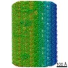
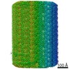
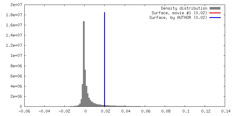
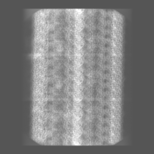
Surface view with section colored by density value
Name: DC1 / type: protein_or_peptide / ID: 34 / Number of copies: 2 / Enantiomer: LEVO EC number: Transferases; Transferring phosphorus-containing groups; Phosphotransferases with an alcohol group as acceptor
pH: 7.4 / Component - Name: HMDEKP Details: 30 mM HEPES, 5 mM MgSO4, 1 mM DTT, 0.5 mM EGTA, 25 mM KCl, PH 7.4
Vitrification
Cryogen name: ETHANE / Chamber humidity: 95 % / Chamber temperature: 277 K / Instrument: FEI VITROBOT MARK IV / Details: blot for 4 seconds before plunging.
-
Electron microscopy
Microscope
FEI TITAN KRIOS
Specialist optics
Spherical aberration corrector: Microscope is equipped with a Cs corrector Energy filter - Name: GIF Bioquantum / Energy filter - Slit width: 20 eV
Image recording
Film or detector model: GATAN K2 QUANTUM (4k x 4k) / Detector mode: COUNTING / Digitization - Dimensions - Width: 3838 pixel / Digitization - Dimensions - Height: 3710 pixel / Digitization - Frames/image: 1-30 / Number grids imaged: 6 / Number real images: 8314 / Average exposure time: 9.0 sec. / Average electron dose: 38.9 e/Å2
Electron beam
Acceleration voltage: 300 kV / Electron source: FIELD EMISSION GUN
In the structure databanks used in Yorodumi, some data are registered as the other names, "COVID-19 virus" and "2019-nCoV". Here are the details of the virus and the list of structure data.
Jan 31, 2019. EMDB accession codes are about to change! (news from PDBe EMDB page)
EMDB accession codes are about to change! (news from PDBe EMDB page)
The allocation of 4 digits for EMDB accession codes will soon come to an end. Whilst these codes will remain in use, new EMDB accession codes will include an additional digit and will expand incrementally as the available range of codes is exhausted. The current 4-digit format prefixed with “EMD-” (i.e. EMD-XXXX) will advance to a 5-digit format (i.e. EMD-XXXXX), and so on. It is currently estimated that the 4-digit codes will be depleted around Spring 2019, at which point the 5-digit format will come into force.
The EM Navigator/Yorodumi systems omit the EMD- prefix.
Related info.:Q: What is EMD? / ID/Accession-code notation in Yorodumi/EM Navigator
Yorodumi is a browser for structure data from EMDB, PDB, SASBDB, etc.
This page is also the successor to EM Navigator detail page, and also detail information page/front-end page for Omokage search.
The word "yorodu" (or yorozu) is an old Japanese word meaning "ten thousand". "mi" (miru) is to see.
Related info.:EMDB / PDB / SASBDB / Comparison of 3 databanks / Yorodumi Search / Aug 31, 2016. New EM Navigator & Yorodumi / Yorodumi Papers / Jmol/JSmol / Function and homology information / Changes in new EM Navigator and Yorodumi
 Movie
Movie Controller

 Yorodumi
 Open data
 Basic information
 Map data
 Sample
 Keywords
 Function and homology information
 Chlamydomonas reinhardtii (plant)
 Authors
 United States, 1 items
  Citation
 Structure visualization
 Downloads & links
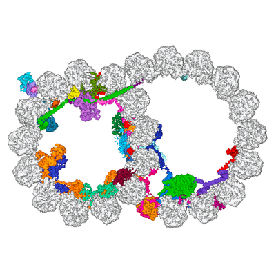 emd_20631.png
 http://ftp.pdbj.org/pub/emdb/structures/EMD-20631
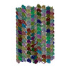


 Z (Sec.)
 Y (Row.)
 X (Col.)


 Sample components

 Processing
 Electron microscopy
 FIELD EMISSION GUN

