 Movie
Movie Controller

[English] 日本語
 Yorodumi
- PDB-6ve7: The inner junction complex of Chlamydomonas reinhardtii doublet m... -
+ Open data

- Basic information
| Entry | Database: PDB / ID: 6ve7 | ||||||||||||
|---|---|---|---|---|---|---|---|---|---|---|---|---|---|
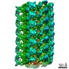
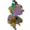
| Title | The inner junction complex of Chlamydomonas reinhardtii doublet microtubule | ||||||||||||
 Components Components |
| ||||||||||||
 Keywords Keywords | STRUCTURAL PROTEIN / cilia / doublet / axoneme / microtubule inner protein | ||||||||||||
| Function / homology |  Function and homology information Function and homology informationaxonemal central pair / axonemal doublet microtubule / positive regulation of cilium-dependent cell motility / regulation of cilium beat frequency involved in ciliary motility / establishment of protein localization to organelle / axoneme assembly / axonemal microtubule / motile cilium / microtubule associated complex / cilium assembly ...axonemal central pair / axonemal doublet microtubule / positive regulation of cilium-dependent cell motility / regulation of cilium beat frequency involved in ciliary motility / establishment of protein localization to organelle / axoneme assembly / axonemal microtubule / motile cilium / microtubule associated complex / cilium assembly / axoneme / microtubule-based process / Hsp70 protein binding / Hsp90 protein binding / structural constituent of cytoskeleton / cytoskeleton / microtubule / Hydrolases; Acting on acid anhydrides; Acting on GTP to facilitate cellular and subcellular movement / calmodulin binding / cilium / ciliary basal body / hydrolase activity / GTPase activity / GTP binding / metal ion binding Similarity search - Function | ||||||||||||
| Biological species |   Chlamydomonas reinhardtii (plant) Chlamydomonas reinhardtii (plant) | ||||||||||||
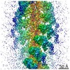
| Method | ELECTRON MICROSCOPY / single particle reconstruction / cryo EM / Resolution: 3.6 Å | ||||||||||||
 Authors Authors | Khalifa, A.A.Z. / Ichikawa, M. / Bui, K.H. | ||||||||||||
| Funding support |  Canada, 3items Canada, 3items
| ||||||||||||
 Citation Citation | Journal: Elife / Year: 2020 Title: The inner junction complex of the cilia is an interaction hub that involves tubulin post-translational modifications. Authors: Ahmad Abdelzaher Zaki Khalifa / Muneyoshi Ichikawa / Daniel Dai / Shintaroh Kubo / Corbin Steven Black / Katya Peri / Thomas S McAlear / Simon Veyron / Shun Kai Yang / Javier Vargas / ...Authors: Ahmad Abdelzaher Zaki Khalifa / Muneyoshi Ichikawa / Daniel Dai / Shintaroh Kubo / Corbin Steven Black / Katya Peri / Thomas S McAlear / Simon Veyron / Shun Kai Yang / Javier Vargas / Susanne Bechstedt / Jean-François Trempe / Khanh Huy Bui /  Abstract: Microtubules are cytoskeletal structures involved in stability, transport and organization in the cell. The building blocks, the α- and β-tubulin heterodimers, form protofilaments that associate ...Microtubules are cytoskeletal structures involved in stability, transport and organization in the cell. The building blocks, the α- and β-tubulin heterodimers, form protofilaments that associate laterally into the hollow microtubule. Microtubule also exists as highly stable doublet microtubules in the cilia where stability is needed for ciliary beating and function. The doublet microtubule maintains its stability through interactions at its inner and outer junctions where its A- and B-tubules meet. Here, using cryo-electron microscopy, bioinformatics and mass spectrometry of the doublets of and , we identified two new inner junction proteins, FAP276 and FAP106, and an inner junction-associated protein, FAP126, thus presenting the complete answer to the inner junction identity and localization. Our structural study of the doublets shows that the inner junction serves as an interaction hub that involves tubulin post-translational modifications. These interactions contribute to the stability of the doublet and hence, normal ciliary motility. | ||||||||||||
| History |
|
- Structure visualization
| Movie |
Movie viewer |
|---|---|
| Structure viewer | Molecule: MolmilJmol/JSmol |
- Downloads & links
-Download
| PDBx/mmCIF format | 6ve7.cif.gz | 4 MB | Display | PDBx/mmCIF format |
|---|---|---|---|---|
| PDB format | pdb6ve7.ent.gz | Display | PDB format | |
| PDBx/mmJSON format | 6ve7.json.gz | Tree view | PDBx/mmJSON format | |
| Others |  Other downloads Other downloads |
-Validation report
| Arichive directory | https://data.pdbj.org/pub/pdb/validation_reports/ve/6ve7ftp://data.pdbj.org/pub/pdb/validation_reports/ve/6ve7 | HTTPS FTP |
|---|
-Related structure data
| Related structure data | 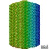 20855MC  20858MC M: map data used to model this data C: citing same article ( |
|---|---|
| Similar structure data |


-Links
 PDBj
PDBj


- Assembly
| Deposited unit | 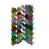
|
|---|---|
| 1 |
|
-Components
-Protein , 8 types, 62 molecules ABCDEFGLMPSXYZefhkmosy01567HIJ...
| #1: Protein | Mass: 27019.803 Da / Num. of mol.: 1 / Source method: isolated from a natural source / Source: (natural) Chlamydomonas reinhardtii (plant) / References: UniProt: A8J098 | ||||||||||||
|---|---|---|---|---|---|---|---|---|---|---|---|---|---|
| #2: Protein | Mass: 68546.508 Da / Num. of mol.: 2 / Source method: isolated from a natural source / Source: (natural) Chlamydomonas reinhardtii (plant) / References: UniProt: A0A2K3D260#3: Protein | Mass: 49638.008 Da / Num. of mol.: 24 / Source method: isolated from a natural source / Source: (natural) Chlamydomonas reinhardtii (plant) / References: UniProt: P09204#4: Protein | | Mass: 15435.397 Da / Num. of mol.: 1 / Source method: isolated from a natural source / Source: (natural) Chlamydomonas reinhardtii (plant) / References: UniProt: A8IVJ1#5: Protein | Mass: 49665.809 Da / Num. of mol.: 24 / Source method: isolated from a natural source / Source: (natural) Chlamydomonas reinhardtii (plant) / References: UniProt: P04690#6: Protein | Mass: 9986.308 Da / Num. of mol.: 2 / Source method: isolated from a natural source / Source: (natural) Chlamydomonas reinhardtii (plant) / References: UniProt: A0A2K3DTN6#7: Protein | Mass: 22193.566 Da / Num. of mol.: 4 / Source method: isolated from a natural source / Source: (natural) Chlamydomonas reinhardtii (plant) / References: UniProt: A8IU92#8: Protein | Mass: 34215.148 Da / Num. of mol.: 4 / Source method: isolated from a natural source / Source: (natural) Chlamydomonas reinhardtii (plant) / References: UniProt: B1B601 |
-Non-polymers , 4 types, 88 molecules 


| #9: Chemical | ChemComp-GTP /  Mass: 523.180 Da / Num. of mol.: 24 / Source method: obtained synthetically / Formula: C10H16N5O14P3 / Comment: GTP, energy-carrying molecule*YM Mass: 523.180 Da / Num. of mol.: 24 / Source method: obtained synthetically / Formula: C10H16N5O14P3 / Comment: GTP, energy-carrying molecule*YM#10: Chemical | ChemComp-MG /  Mass: 24.305 Da / Num. of mol.: 24 / Source method: obtained synthetically / Formula: Mg Mass: 24.305 Da / Num. of mol.: 24 / Source method: obtained synthetically / Formula: Mg#11: Chemical | ChemComp-GDP /  Type: RNA linking / Mass: 443.201 Da / Num. of mol.: 24 / Source method: obtained synthetically / Formula: C10H15N5O11P2 / Comment: GDP, energy-carrying molecule*YM Type: RNA linking / Mass: 443.201 Da / Num. of mol.: 24 / Source method: obtained synthetically / Formula: C10H15N5O11P2 / Comment: GDP, energy-carrying molecule*YM#12: Chemical | ChemComp-TA1 /  Mass: 853.906 Da / Num. of mol.: 16 / Source method: obtained synthetically / Formula: C47H51NO14 / Comment: medication*YM Mass: 853.906 Da / Num. of mol.: 16 / Source method: obtained synthetically / Formula: C47H51NO14 / Comment: medication*YM |
|---|
-Details
| Has ligand of interest | N |
|---|---|
| Has protein modification | Y |
-Experimental details
-Experiment
| Experiment | Method: ELECTRON MICROSCOPY |
|---|---|
| EM experiment | Aggregation state: FILAMENT / 3D reconstruction method: single particle reconstruction |
- Sample preparation
| Component | Name: The inner junction complex of Chlamydomonas axoneme / Type: ORGANELLE OR CELLULAR COMPONENT / Entity ID: #1-#8 / Source: NATURAL |
|---|---|
| Molecular weight | Experimental value: NO |
| Source (natural) | Organism: Chlamydomonas reinhardtii (plant) / Strain: CC-124 / Organelle: cilia |
| Buffer solution | pH: 7.4 |
| Specimen | Conc.: 2 mg/ml / Embedding applied: NO / Shadowing applied: NO / Staining applied: NO / Vitrification applied: YES |
| Specimen support | Grid material: COPPER / Grid mesh size: 200 divisions/in. / Grid type: Quantifoil R2/2 |
| Vitrification | Instrument: FEI VITROBOT MARK IV / Cryogen name: ETHANE / Humidity: 100 % / Chamber temperature: 298 K |
- Electron microscopy imaging
| Experimental equipment |  Model: Titan Krios / Image courtesy: FEI Company |
|---|---|
| Microscopy | Model: FEI TITAN KRIOS |
| Electron gun | Electron source:  FIELD EMISSION GUN / Accelerating voltage: 300 kV / Illumination mode: FLOOD BEAM FIELD EMISSION GUN / Accelerating voltage: 300 kV / Illumination mode: FLOOD BEAM |
| Electron lens | Mode: BRIGHT FIELD / Nominal magnification: 59000 X / Nominal defocus max: 4000 nm / Nominal defocus min: 1000 nm / Calibrated defocus min: 1000 nm / Calibrated defocus max: 4000 nm / Cs: 2.7 mm / C2 aperture diameter: 100 µm / Alignment procedure: COMA FREE |
| Specimen holder | Cryogen: NITROGEN / Specimen holder model: FEI TITAN KRIOS AUTOGRID HOLDER |
| Image recording | Average exposure time: 2 sec. / Electron dose: 45 e/Å2 / Detector mode: INTEGRATING / Film or detector model: FEI FALCON II (4k x 4k) / Num. of grids imaged: 3 / Num. of real images: 9528 |
| Image scans | Sampling size: 14 µm / Width: 4096 / Height: 4096 / Movie frames/image: 7 / Used frames/image: 1-7 |
- Processing
| EM software |
| ||||||||||||||||||||||||||||||||||||||||||||||||||
|---|---|---|---|---|---|---|---|---|---|---|---|---|---|---|---|---|---|---|---|---|---|---|---|---|---|---|---|---|---|---|---|---|---|---|---|---|---|---|---|---|---|---|---|---|---|---|---|---|---|---|---|
| CTF correction | Type: PHASE FLIPPING AND AMPLITUDE CORRECTION | ||||||||||||||||||||||||||||||||||||||||||||||||||
| Particle selection | Num. of particles selected: 270713 | ||||||||||||||||||||||||||||||||||||||||||||||||||
| Symmetry | Point symmetry: C1 (asymmetric) | ||||||||||||||||||||||||||||||||||||||||||||||||||
| 3D reconstruction | Resolution: 3.6 Å / Resolution method: FSC 0.143 CUT-OFF / Num. of particles: 270713 / Algorithm: BACK PROJECTION / Num. of class averages: 1 / Symmetry type: POINT | ||||||||||||||||||||||||||||||||||||||||||||||||||
| Atomic model building | Protocol: AB INITIO MODEL |
