Movie
Movie Controller

[English] 日本語
 Yorodumi
- EMDB-4531: Cryo-EM structure of the 50S ribosomal subunit at 2.83 Angstroms ... -
+ Open data

- Basic information
| Entry | Database: EMDB / ID: EMD-4531 | |||||||||
|---|---|---|---|---|---|---|---|---|---|---|
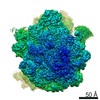
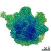
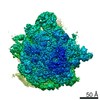
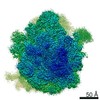
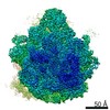
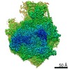
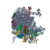
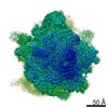
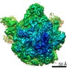
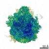
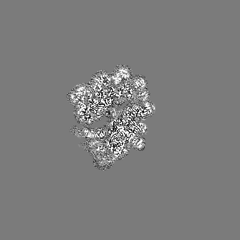
| Title | Cryo-EM structure of the 50S ribosomal subunit at 2.83 Angstroms with modeled GBC SecM peptide | |||||||||
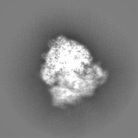
 Map data Map data | postprocessed map | |||||||||
 Sample Sample |
| |||||||||
 Keywords Keywords | Translation / 50S ribosome / Nascent peptide chain / RIBOSOME | |||||||||
| Function / homology |  Function and homology information Function and homology informationstructural constituent of eye lens / lens development in camera-type eye / transcriptional attenuation / endoribonuclease inhibitor activity / positive regulation of ribosome biogenesis / RNA-binding transcription regulator activity / negative regulation of cytoplasmic translation / DnaA-L2 complex / translation repressor activity / visual perception ...structural constituent of eye lens / lens development in camera-type eye / transcriptional attenuation / endoribonuclease inhibitor activity / positive regulation of ribosome biogenesis / RNA-binding transcription regulator activity / negative regulation of cytoplasmic translation / DnaA-L2 complex / translation repressor activity / visual perception / negative regulation of DNA-templated DNA replication initiation / mRNA regulatory element binding translation repressor activity / response to reactive oxygen species / cytosolic ribosome assembly / ribosome assembly / assembly of large subunit precursor of preribosome / regulation of cell growth / DNA-templated transcription termination / response to radiation / mRNA 5'-UTR binding / large ribosomal subunit / transferase activity / ribosome binding / 5S rRNA binding / ribosomal large subunit assembly / large ribosomal subunit rRNA binding / cytosolic large ribosomal subunit / cytoplasmic translation / tRNA binding / negative regulation of translation / rRNA binding / structural constituent of ribosome / ribosome / translation / ribonucleoprotein complex / response to antibiotic / negative regulation of DNA-templated transcription / mRNA binding / DNA binding / RNA binding / zinc ion binding / cytoplasm / cytosol Similarity search - Function | |||||||||
| Biological species |   | |||||||||
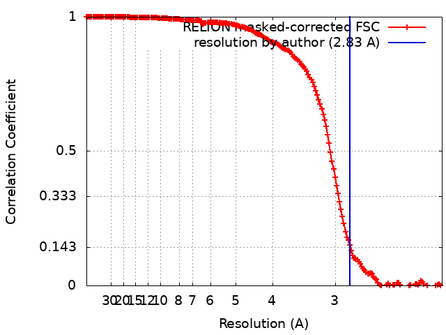
| Method | single particle reconstruction / cryo EM / Resolution: 2.83 Å | |||||||||
 Authors Authors | Schulte L / Reitz J | |||||||||
 Citation Citation | Journal: Nat Commun / Year: 2020 Title: Cysteine oxidation and disulfide formation in the ribosomal exit tunnel. Authors: Linda Schulte / Jiafei Mao / Julian Reitz / Sridhar Sreeramulu / Denis Kudlinzki / Victor-Valentin Hodirnau / Jakob Meier-Credo / Krishna Saxena / Florian Buhr / Julian D Langer / Martin ...Authors: Linda Schulte / Jiafei Mao / Julian Reitz / Sridhar Sreeramulu / Denis Kudlinzki / Victor-Valentin Hodirnau / Jakob Meier-Credo / Krishna Saxena / Florian Buhr / Julian D Langer / Martin Blackledge / Achilleas S Frangakis / Clemens Glaubitz / Harald Schwalbe /     Abstract: Understanding the conformational sampling of translation-arrested ribosome nascent chain complexes is key to understand co-translational folding. Up to now, coupling of cysteine oxidation, disulfide ...Understanding the conformational sampling of translation-arrested ribosome nascent chain complexes is key to understand co-translational folding. Up to now, coupling of cysteine oxidation, disulfide bond formation and structure formation in nascent chains has remained elusive. Here, we investigate the eye-lens protein γB-crystallin in the ribosomal exit tunnel. Using mass spectrometry, theoretical simulations, dynamic nuclear polarization-enhanced solid-state nuclear magnetic resonance and cryo-electron microscopy, we show that thiol groups of cysteine residues undergo S-glutathionylation and S-nitrosylation and form non-native disulfide bonds. Thus, covalent modification chemistry occurs already prior to nascent chain release as the ribosome exit tunnel provides sufficient space even for disulfide bond formation which can guide protein folding. | |||||||||
| History |
|
- Structure visualization
| Movie |
Movie viewer |
|---|---|
| Structure viewer | EM map: SurfViewMolmilJmol/JSmol |
| Supplemental images |

- Downloads & links
-EMDB archive
| Map data | emd_4531.map.gz | 395.9 MB | EMDB map data format | |
|---|---|---|---|---|
| Header (meta data) | emd-4531-v30.xmlemd-4531.xml | 58 KB 58 KB | Display Display | EMDB header |
| FSC (resolution estimation) | emd_4531_fsc.xml | 16.9 KB | Display | FSC data file |
| Images | 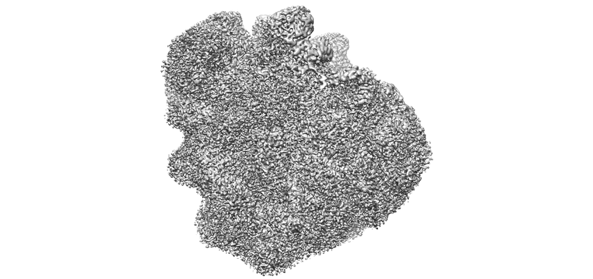 emd_4531.png emd_4531.png | 104.1 KB | ||
| Filedesc metadata | emd-4531.cif.gz | 11 KB | ||
| Others | emd_4531_additional.map.gzemd_4531_half_map_1.map.gzemd_4531_half_map_2.map.gz | 30.9 MB 338.2 MB 338.3 MB | ||
| Archive directory |  http://ftp.pdbj.org/pub/emdb/structures/EMD-4531ftp://ftp.pdbj.org/pub/emdb/structures/EMD-4531 http://ftp.pdbj.org/pub/emdb/structures/EMD-4531ftp://ftp.pdbj.org/pub/emdb/structures/EMD-4531 | HTTPS FTP |
-Related structure data
| Related structure data |  6qdwMC  6ys3C M: atomic model generated by this map C: citing same article ( |
|---|---|
| Similar structure data |

-Links
| EMDB pages | EMDB (EBI/PDBe) / EMDataResource |
|---|---|
| Related items in Molecule of the Month |


-Map
| File | Download / File: emd_4531.map.gz / Format: CCP4 / Size: 421.9 MB / Type: IMAGE STORED AS FLOATING POINT NUMBER (4 BYTES) | ||||||||||||||||||||||||||||||||||||||||||||||||||||||||||||
|---|---|---|---|---|---|---|---|---|---|---|---|---|---|---|---|---|---|---|---|---|---|---|---|---|---|---|---|---|---|---|---|---|---|---|---|---|---|---|---|---|---|---|---|---|---|---|---|---|---|---|---|---|---|---|---|---|---|---|---|---|---|
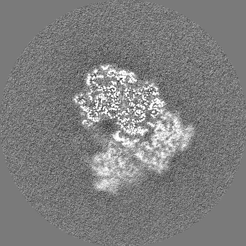
| Annotation | postprocessed map | ||||||||||||||||||||||||||||||||||||||||||||||||||||||||||||
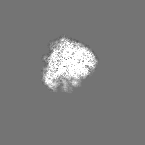
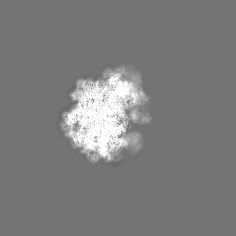
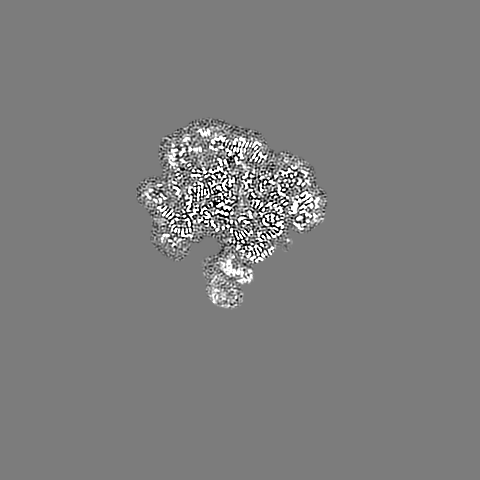
| Projections & slices | Image control
Images are generated by Spider. | ||||||||||||||||||||||||||||||||||||||||||||||||||||||||||||
| Voxel size | X=Y=Z: 1.05 Å | ||||||||||||||||||||||||||||||||||||||||||||||||||||||||||||
| Density |
| ||||||||||||||||||||||||||||||||||||||||||||||||||||||||||||
| Symmetry | Space group: 1 | ||||||||||||||||||||||||||||||||||||||||||||||||||||||||||||
| Details | EMDB XML:
CCP4 map header:
| ||||||||||||||||||||||||||||||||||||||||||||||||||||||||||||
 Z (Sec.)
 Y (Row.)
 X (Col.)


-Supplemental data
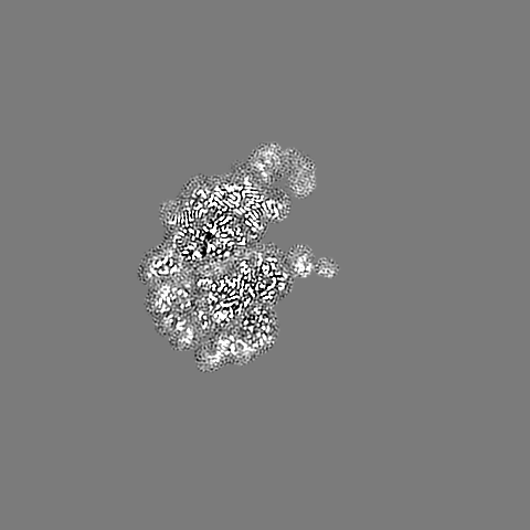
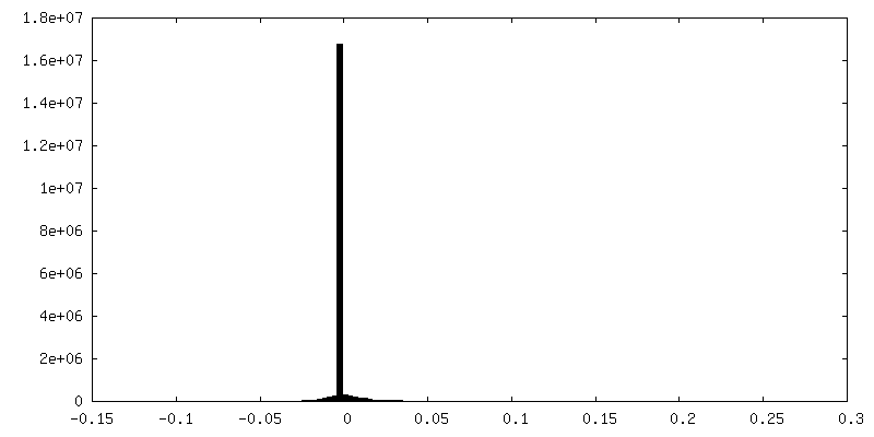
-Additional map: masked postprocessed map
| File | emd_4531_additional.map | ||||||||||||
|---|---|---|---|---|---|---|---|---|---|---|---|---|---|
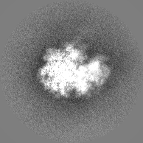
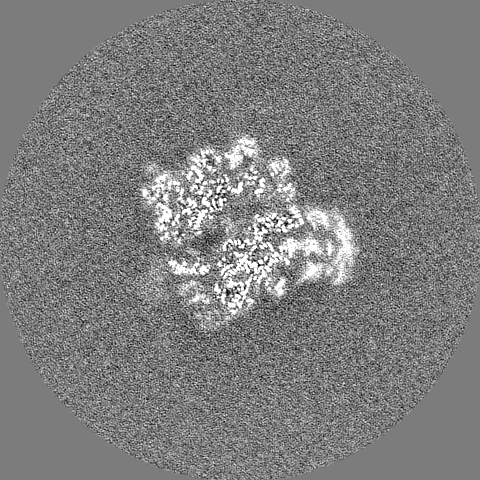
| Annotation | masked postprocessed map | ||||||||||||
| Projections & Slices |
| ||||||||||||
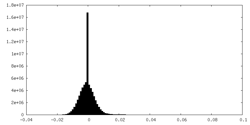
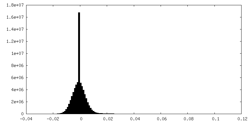
| Density Histograms |

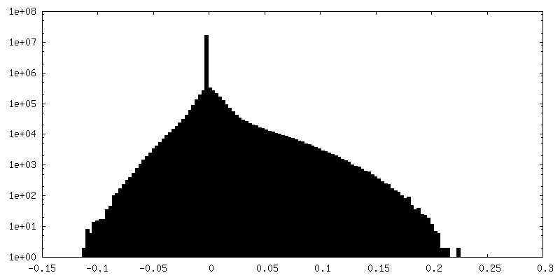
-Half map: EM Half map 2
| File | emd_4531_half_map_1.map | ||||||||||||
|---|---|---|---|---|---|---|---|---|---|---|---|---|---|
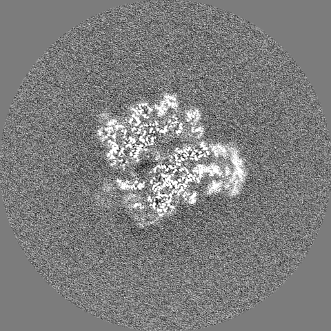
| Annotation | EM Half map 2 | ||||||||||||
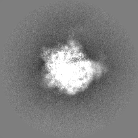
| Projections & Slices |
| ||||||||||||
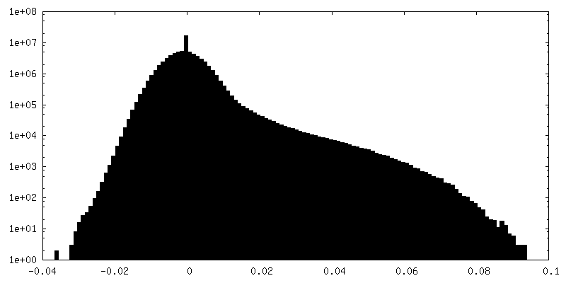
| Density Histograms |


-Half map: EM Half map 1
| File | emd_4531_half_map_2.map | ||||||||||||
|---|---|---|---|---|---|---|---|---|---|---|---|---|---|
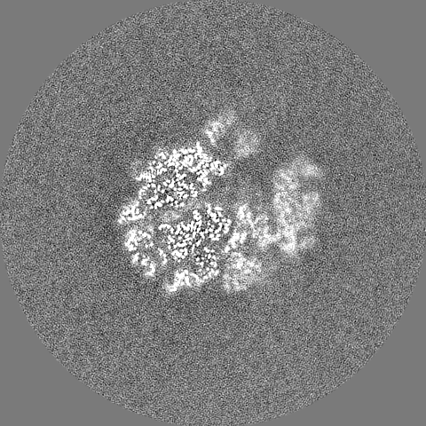
| Annotation | EM Half map 1 | ||||||||||||
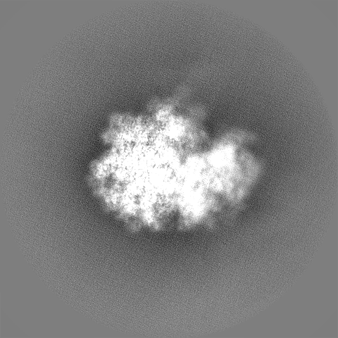
| Projections & Slices |
| ||||||||||||
| Density Histograms |


- Sample components
+Entire : 50S ribosomal + GBC SecM peptide complex
+Supramolecule #1: 50S ribosomal + GBC SecM peptide complex
+Supramolecule #2: GBC SecM peptide
+Supramolecule #3: 50 ribosomal subunit
+Macromolecule #1: 50S ribosomal protein L28
+Macromolecule #2: 50S ribosomal protein L29
+Macromolecule #3: 50S ribosomal protein L30
+Macromolecule #4: 50S ribosomal protein L32
+Macromolecule #5: 50S ribosomal protein L33
+Macromolecule #6: 50S ribosomal protein L34
+Macromolecule #7: 50S ribosomal protein L35
+Macromolecule #8: 50S ribosomal protein L36
+Macromolecule #11: 50S ribosomal protein L2
+Macromolecule #12: 50S ribosomal protein L3
+Macromolecule #13: 50S ribosomal protein L4
+Macromolecule #14: 50S ribosomal protein L5
+Macromolecule #15: 50S ribosomal protein L6
+Macromolecule #16: 50S ribosomal protein L9
+Macromolecule #17: 50S ribosomal protein L13
+Macromolecule #18: 50S ribosomal protein L14
+Macromolecule #19: 50S ribosomal protein L15
+Macromolecule #20: 50S ribosomal protein L16
+Macromolecule #21: 50S ribosomal protein L17
+Macromolecule #22: 50S ribosomal protein L18
+Macromolecule #23: 50S ribosomal protein L19
+Macromolecule #24: 50S ribosomal protein L20
+Macromolecule #25: 50S ribosomal protein L21
+Macromolecule #26: 50S ribosomal protein L22
+Macromolecule #27: 50S ribosomal protein L23
+Macromolecule #28: 50S ribosomal protein L24
+Macromolecule #30: 50S ribosomal protein L25
+Macromolecule #31: 50S ribosomal protein L27
+Macromolecule #32: Gamma-crystallin B
+Macromolecule #9: 5S rRNA
+Macromolecule #10: 23S rRNA
+Macromolecule #29: glycine-tRNA glyT
-Experimental details
-Structure determination
| Method | cryo EM |
|---|---|
 Processing Processing | single particle reconstruction |
| Aggregation state | particle |
-Sample preparation
| Concentration | 0.12 mg/mL | |||||||||||||||
|---|---|---|---|---|---|---|---|---|---|---|---|---|---|---|---|---|
| Buffer | pH: 7.5 Component:
Details: Tico Buffer | |||||||||||||||
| Grid | Model: Quantifoil R2/1 / Material: COPPER / Mesh: 300 / Support film - Material: CARBON / Support film - topology: CONTINUOUS / Support film - Film thickness: 5 / Pretreatment - Type: GLOW DISCHARGE / Pretreatment - Time: 20 sec. | |||||||||||||||
| Vitrification | Cryogen name: ETHANE / Chamber humidity: 100 % / Chamber temperature: 277.15 K / Instrument: FEI VITROBOT MARK IV |
- Electron microscopy
| Microscope | FEI TITAN KRIOS |
|---|---|
| Specialist optics | Energy filter - Name: GIF Quantum SE |
| Image recording | Film or detector model: GATAN K2 SUMMIT (4k x 4k) / Detector mode: COUNTING / Number grids imaged: 3 / Average exposure time: 0.2 sec. / Average electron dose: 1.66 e/Å2 |
| Electron beam | Acceleration voltage: 300 kV / Electron source:  FIELD EMISSION GUN FIELD EMISSION GUN |
| Electron optics | Illumination mode: FLOOD BEAM / Imaging mode: BRIGHT FIELD / Cs: 2.7 mm |
| Sample stage | Specimen holder model: FEI TITAN KRIOS AUTOGRID HOLDER / Cooling holder cryogen: NITROGEN |
| Experimental equipment |  Model: Titan Krios / Image courtesy: FEI Company |