Movie
Movie Controller

[English] 日本語
 Yorodumi
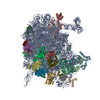
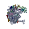
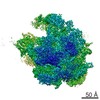
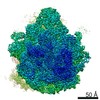
- EMDB-10977: The structure of the Atp25 bound assembly intermediate of the mit... -
+ Open data

- Basic information
| Entry | Database: EMDB / ID: EMD-10977 | |||||||||||||||
|---|---|---|---|---|---|---|---|---|---|---|---|---|---|---|---|---|
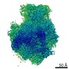
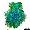
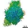
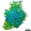
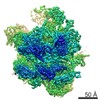
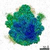
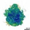
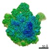
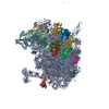
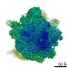
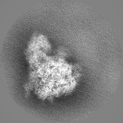
| Title | The structure of the Atp25 bound assembly intermediate of the mitoribosome from Neurospora crassa | |||||||||||||||
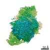
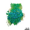
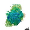
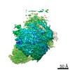
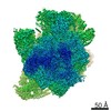
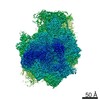
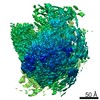
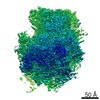
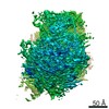
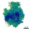
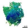
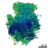
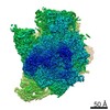
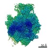
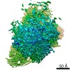
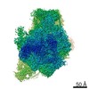
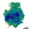
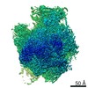
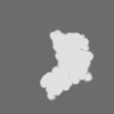
 Map data Map data | Consensus map for the assembly intermediate with bound Atp25 | |||||||||||||||
 Sample Sample |
| |||||||||||||||
 Keywords Keywords | Neurospora crassa / translating Mitoribosomes / tRNA / mRNA / mL108 / TRANSLATION | |||||||||||||||
| Function / homology |  Function and homology information Function and homology informationmitochondrial gene expression / ribonuclease III activity / DNA strand exchange activity / mitochondrial large ribosomal subunit / mitochondrial translation / mRNA stabilization / RNA processing / double-stranded RNA binding / single-stranded DNA binding / large ribosomal subunit ...mitochondrial gene expression / ribonuclease III activity / DNA strand exchange activity / mitochondrial large ribosomal subunit / mitochondrial translation / mRNA stabilization / RNA processing / double-stranded RNA binding / single-stranded DNA binding / large ribosomal subunit / transferase activity / large ribosomal subunit rRNA binding / mitochondrial inner membrane / negative regulation of translation / rRNA binding / structural constituent of ribosome / ribosome / translation / ribonucleoprotein complex / mRNA binding / mitochondrion / RNA binding / metal ion binding Similarity search - Function | |||||||||||||||
| Biological species |  Neurospora crassa OR74A (fungus) Neurospora crassa OR74A (fungus) | |||||||||||||||
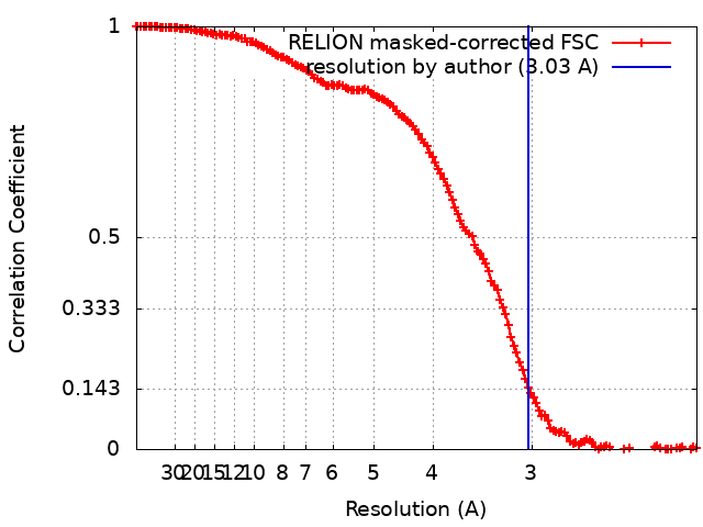
| Method | single particle reconstruction / cryo EM / Resolution: 3.03 Å | |||||||||||||||
 Authors Authors | Amunts A / Itoh Y | |||||||||||||||
| Funding support |  Sweden, European Union, 4 items Sweden, European Union, 4 items
| |||||||||||||||
 Citation Citation | Journal: Nat Commun / Year: 2020 Title: Analysis of translating mitoribosome reveals functional characteristics of translation in mitochondria of fungi. Authors: Yuzuru Itoh / Andreas Naschberger / Narges Mortezaei / Johannes M Herrmann / Alexey Amunts /  Abstract: Mitoribosomes are specialized protein synthesis machineries in mitochondria. However, how mRNA binds to its dedicated channel, and tRNA moves as the mitoribosomal subunit rotate with respect to each ...Mitoribosomes are specialized protein synthesis machineries in mitochondria. However, how mRNA binds to its dedicated channel, and tRNA moves as the mitoribosomal subunit rotate with respect to each other is not understood. We report models of the translating fungal mitoribosome with mRNA, tRNA and nascent polypeptide, as well as an assembly intermediate. Nicotinamide adenine dinucleotide (NAD) is found in the central protuberance of the large subunit, and the ATPase inhibitory factor 1 (IF) in the small subunit. The models of the active mitoribosome explain how mRNA binds through a dedicated protein platform on the small subunit, tRNA is translocated with the help of the protein mL108, bridging it with L1 stalk on the large subunit, and nascent polypeptide paths through a newly shaped exit tunnel involving a series of structural rearrangements. An assembly intermediate is modeled with the maturation factor Atp25, providing insight into the biogenesis of the mitoribosomal large subunit and translation regulation. | |||||||||||||||
| History |
|
- Structure visualization
| Movie |
Movie viewer |
|---|---|
| Structure viewer | EM map: SurfViewMolmilJmol/JSmol |
| Supplemental images |

- Downloads & links
-EMDB archive
| Map data | emd_10977.map.gz | 142.8 MB | EMDB map data format | |
|---|---|---|---|---|
| Header (meta data) | emd-10977-v30.xmlemd-10977.xml | 69.4 KB 69.4 KB | Display Display | EMDB header |
| FSC (resolution estimation) | emd_10977_fsc.xml | 14.2 KB | Display | FSC data file |
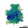
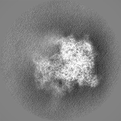
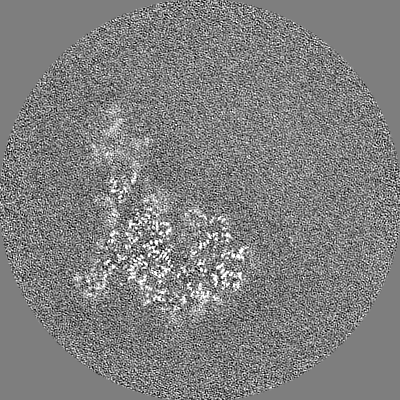
| Images | 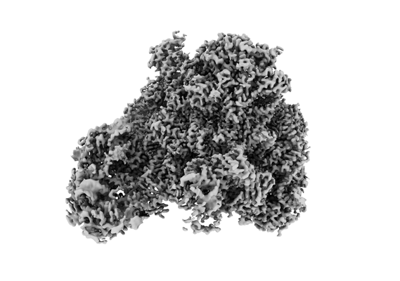 emd_10977.png emd_10977.png | 74.7 KB | ||
| Masks | emd_10977_msk_1.map | 244.1 MB | Mask map | |
| Filedesc metadata | emd-10977.cif.gz | 16.5 KB | ||
| Others | emd_10977_half_map_1.map.gzemd_10977_half_map_2.map.gz | 195.3 MB 195.4 MB | ||
| Archive directory |  http://ftp.pdbj.org/pub/emdb/structures/EMD-10977ftp://ftp.pdbj.org/pub/emdb/structures/EMD-10977 http://ftp.pdbj.org/pub/emdb/structures/EMD-10977ftp://ftp.pdbj.org/pub/emdb/structures/EMD-10977 | HTTPS FTP |
-Related structure data
| Related structure data | 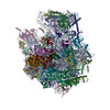 6ywvMC 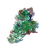 6yw5C 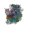 6yweC 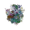 6ywsC  6ywxC 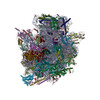 6ywyC M: atomic model generated by this map C: citing same article ( |
|---|---|
| Similar structure data |


-Links
| EMDB pages | EMDB (EBI/PDBe) / EMDataResource |
|---|---|
| Related items in Molecule of the Month |


-Map
| File | Download / File: emd_10977.map.gz / Format: CCP4 / Size: 244.1 MB / Type: IMAGE STORED AS FLOATING POINT NUMBER (4 BYTES) | ||||||||||||||||||||||||||||||||||||||||||||||||||||||||||||||||||||
|---|---|---|---|---|---|---|---|---|---|---|---|---|---|---|---|---|---|---|---|---|---|---|---|---|---|---|---|---|---|---|---|---|---|---|---|---|---|---|---|---|---|---|---|---|---|---|---|---|---|---|---|---|---|---|---|---|---|---|---|---|---|---|---|---|---|---|---|---|---|
| Annotation | Consensus map for the assembly intermediate with bound Atp25 | ||||||||||||||||||||||||||||||||||||||||||||||||||||||||||||||||||||
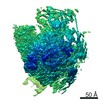
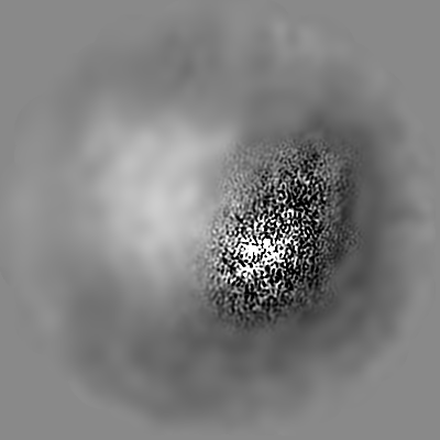
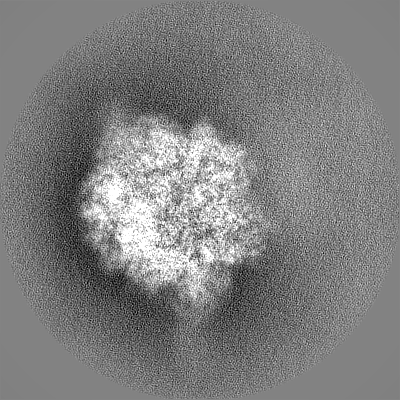
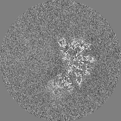
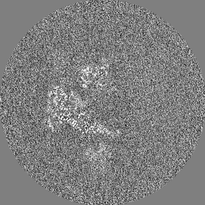
| Projections & slices | Image control
Images are generated by Spider. | ||||||||||||||||||||||||||||||||||||||||||||||||||||||||||||||||||||
| Voxel size | X=Y=Z: 1.06 Å | ||||||||||||||||||||||||||||||||||||||||||||||||||||||||||||||||||||
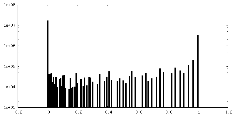
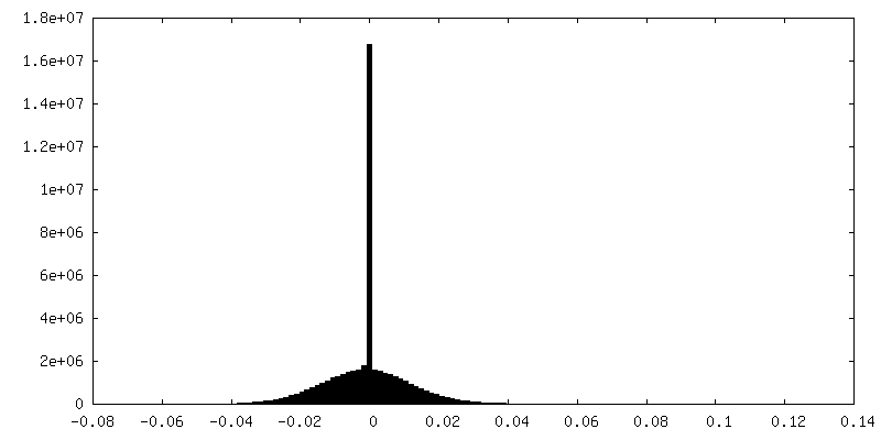
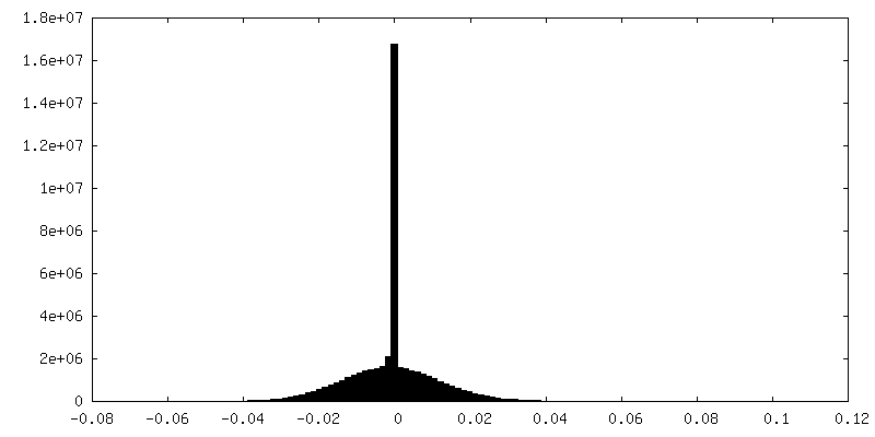
| Density |
| ||||||||||||||||||||||||||||||||||||||||||||||||||||||||||||||||||||
| Symmetry | Space group: 1 | ||||||||||||||||||||||||||||||||||||||||||||||||||||||||||||||||||||
| Details | EMDB XML:
CCP4 map header:
| ||||||||||||||||||||||||||||||||||||||||||||||||||||||||||||||||||||
 Z (Sec.)
 Y (Row.)
 X (Col.)


-Supplemental data
-Mask #1
| File | emd_10977_msk_1.map | ||||||||||||
|---|---|---|---|---|---|---|---|---|---|---|---|---|---|
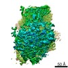
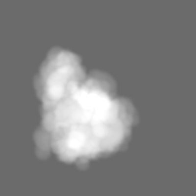
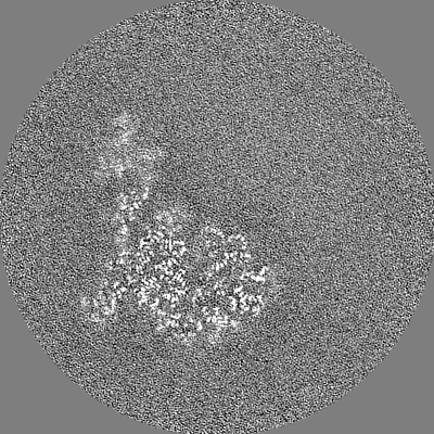
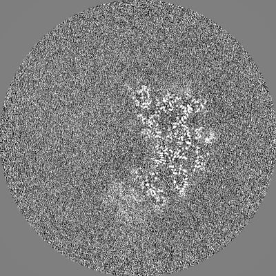
| Projections & Slices |
| ||||||||||||
| Density Histograms |


-Half map: #1
| File | emd_10977_half_map_1.map | ||||||||||||
|---|---|---|---|---|---|---|---|---|---|---|---|---|---|
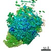
| Projections & Slices |
| ||||||||||||
| Density Histograms |


-Half map: #2
| File | emd_10977_half_map_2.map | ||||||||||||
|---|---|---|---|---|---|---|---|---|---|---|---|---|---|
| Projections & Slices |
| ||||||||||||
| Density Histograms |

- Sample components
+Entire : Assembly intermediate of the mitoribosome of Neurospora crassa
+Supramolecule #1: Assembly intermediate of the mitoribosome of Neurospora crassa
+Macromolecule #1: 23 S rRNA
+Macromolecule #2: 60S ribosomal protein L2
+Macromolecule #3: 60S ribosomal protein L3
+Macromolecule #4: 60S ribosomal protein L4, variant
+Macromolecule #5: 50S ribosomal protein L5
+Macromolecule #6: 60S ribosomal protein L6
+Macromolecule #7: Uncharacterized protein
+Macromolecule #8: 60S ribosomal protein L19
+Macromolecule #9: Ribosomal protein L13
+Macromolecule #10: 50S ribosomal protein L14
+Macromolecule #11: 50S ribosomal subunit protein L15
+Macromolecule #12: 60S ribosomal protein L16
+Macromolecule #13: 50S ribosomal protein L17
+Macromolecule #14: Mitochondrial ribosomal protein
+Macromolecule #15: Aconitate hydratase
+Macromolecule #16: Mitochondrial large ribosomal subunit
+Macromolecule #17: Mitochondrial ribosomal protein subunit L23
+Macromolecule #18: KOW domain-containing protein
+Macromolecule #19: Uncharacterized protein
+Macromolecule #20: 50S ribosomal protein L24
+Macromolecule #21: 54S ribosomal protein L4, mitochondrial
+Macromolecule #22: 50S ribosomal protein L30
+Macromolecule #23: Uncharacterized protein
+Macromolecule #24: Mitochondrial ribosomal protein subunit L32
+Macromolecule #25: Uncharacterized protein
+Macromolecule #26: Uncharacterized protein
+Macromolecule #27: Ribosomal protein
+Macromolecule #28: Mitochondrial large ribosomal subunit YmL35
+Macromolecule #29: Uncharacterized protein
+Macromolecule #30: Uncharacterized protein
+Macromolecule #31: Mitochondrial ribosomal protein L43
+Macromolecule #32: 60S ribosomal protein L3
+Macromolecule #33: 50S ribosomal subunit L30
+Macromolecule #34: Uncharacterized protein
+Macromolecule #35: Uncharacterized protein
+Macromolecule #36: Mitochondrial ribosomal protein L44
+Macromolecule #37: Uncharacterized protein
+Macromolecule #38: RNase III domain-containing protein
+Macromolecule #39: 60S ribosomal protein L20
+Macromolecule #40: Mitochondrial 60S ribosomal protein L25
+Macromolecule #41: 54S ribosomal protein L31, mitochondrial
+Macromolecule #42: Uncharacterized protein
+Macromolecule #43: ATPase synthesis protein 25, mitochondrial
+Macromolecule #44: MAGNESIUM ION
+Macromolecule #45: NICOTINAMIDE-ADENINE-DINUCLEOTIDE

+Macromolecule #46: SPERMINE

+Macromolecule #47: POTASSIUM ION
+Macromolecule #48: ZINC ION
-Experimental details
-Structure determination
| Method | cryo EM |
|---|---|
 Processing Processing | single particle reconstruction |
| Aggregation state | particle |
-Sample preparation
| Buffer | pH: 7.5 Component:
| |||||||||||||||||||||
|---|---|---|---|---|---|---|---|---|---|---|---|---|---|---|---|---|---|---|---|---|---|---|
| Grid | Model: Quantifoil R2/2 / Material: COPPER / Support film - Material: CARBON / Support film - topology: CONTINUOUS / Support film - Film thickness: 2.7 | |||||||||||||||||||||
| Vitrification | Cryogen name: ETHANE / Chamber humidity: 100 % / Chamber temperature: 277 K / Instrument: FEI VITROBOT MARK IV |
- Electron microscopy
| Microscope | FEI TITAN KRIOS |
|---|---|
| Specialist optics | Energy filter - Name: GIF Quantum LS / Energy filter - Slit width: 20 eV |
| Image recording | Film or detector model: GATAN K2 SUMMIT (4k x 4k) / Detector mode: COUNTING / Digitization - Frames/image: 1-20 / Number real images: 3172 / Average electron dose: 35.0 e/Å2 |
| Electron beam | Acceleration voltage: 300 kV / Electron source:  FIELD EMISSION GUN FIELD EMISSION GUN |
| Electron optics | C2 aperture diameter: 70.0 µm / Calibrated magnification: 130000 / Illumination mode: FLOOD BEAM / Imaging mode: BRIGHT FIELD |
| Sample stage | Specimen holder model: FEI TITAN KRIOS AUTOGRID HOLDER / Cooling holder cryogen: NITROGEN |
| Experimental equipment |  Model: Titan Krios / Image courtesy: FEI Company |