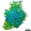
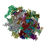
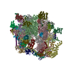
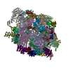
 Movie
Movie Controller

[English] 日本語
 Yorodumi
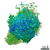
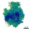
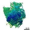
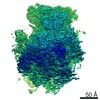
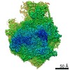
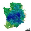
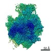
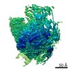
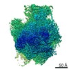
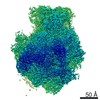
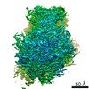
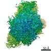
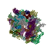
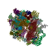
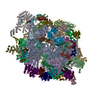
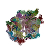
- PDB-6ywy: The structure of the mitoribosome from Neurospora crassa with bou... -
+ Open data

- Basic information
| Entry | Database: PDB / ID: 6ywy | |||||||||||||||
|---|---|---|---|---|---|---|---|---|---|---|---|---|---|---|---|---|
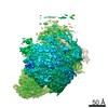
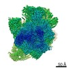
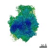
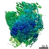
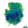
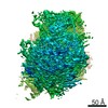
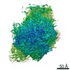
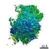
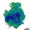
| Title | The structure of the mitoribosome from Neurospora crassa with bound tRNA at the P-site | |||||||||||||||
 Components Components |
| |||||||||||||||
 Keywords Keywords | TRANSLATION / Neurospora crassa / translating Mitoribosomes / tRNA / mRNA / mL108 | |||||||||||||||
| Function / homology |  Function and homology information Function and homology information: / 3-hydroxyisobutyryl-CoA hydrolase / 3-hydroxyisobutyryl-CoA hydrolase activity / L-valine catabolic process / negative regulation of ATP-dependent activity / ATPase inhibitor activity / ribonuclease III activity / DNA strand exchange activity / mitochondrial large ribosomal subunit / mitochondrial ribosome ...: / 3-hydroxyisobutyryl-CoA hydrolase / 3-hydroxyisobutyryl-CoA hydrolase activity / L-valine catabolic process / negative regulation of ATP-dependent activity / ATPase inhibitor activity / ribonuclease III activity / DNA strand exchange activity / mitochondrial large ribosomal subunit / mitochondrial ribosome / mitochondrial small ribosomal subunit / mitochondrial translation / superoxide dismutase activity / RNA processing / rRNA processing / peroxisome / double-stranded RNA binding / single-stranded DNA binding / large ribosomal subunit / transferase activity / small ribosomal subunit / small ribosomal subunit rRNA binding / rRNA binding / structural constituent of ribosome / ribosome / translation / ribonucleoprotein complex / mitochondrion / RNA binding / metal ion binding / cytoplasm Similarity search - Function | |||||||||||||||
| Biological species |  Neurospora crassa (fungus) Neurospora crassa (fungus) | |||||||||||||||
| Method | ELECTRON MICROSCOPY / single particle reconstruction / cryo EM / Resolution: 3.05 Å | |||||||||||||||
 Authors Authors | Amunts, A. / Itoh, Y. / Naschberger, A. | |||||||||||||||
| Funding support |  Sweden, European Union, 4items Sweden, European Union, 4items
| |||||||||||||||
 Citation Citation | Journal: Nat Commun / Year: 2020 Title: Analysis of translating mitoribosome reveals functional characteristics of translation in mitochondria of fungi. Authors: Yuzuru Itoh / Andreas Naschberger / Narges Mortezaei / Johannes M Herrmann / Alexey Amunts /  Abstract: Mitoribosomes are specialized protein synthesis machineries in mitochondria. However, how mRNA binds to its dedicated channel, and tRNA moves as the mitoribosomal subunit rotate with respect to each ...Mitoribosomes are specialized protein synthesis machineries in mitochondria. However, how mRNA binds to its dedicated channel, and tRNA moves as the mitoribosomal subunit rotate with respect to each other is not understood. We report models of the translating fungal mitoribosome with mRNA, tRNA and nascent polypeptide, as well as an assembly intermediate. Nicotinamide adenine dinucleotide (NAD) is found in the central protuberance of the large subunit, and the ATPase inhibitory factor 1 (IF) in the small subunit. The models of the active mitoribosome explain how mRNA binds through a dedicated protein platform on the small subunit, tRNA is translocated with the help of the protein mL108, bridging it with L1 stalk on the large subunit, and nascent polypeptide paths through a newly shaped exit tunnel involving a series of structural rearrangements. An assembly intermediate is modeled with the maturation factor Atp25, providing insight into the biogenesis of the mitoribosomal large subunit and translation regulation. | |||||||||||||||
| History |
|
- Structure visualization
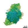
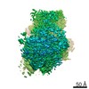
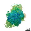
| Movie |
Movie viewer |
|---|---|
| Structure viewer | Molecule: MolmilJmol/JSmol |
- Downloads & links
-Download
| PDBx/mmCIF format | 6ywy.cif.gz | 9.6 MB | Display | PDBx/mmCIF format |
|---|---|---|---|---|
| PDB format | pdb6ywy.ent.gz | Display | PDB format | |
| PDBx/mmJSON format | 6ywy.json.gz | Tree view | PDBx/mmJSON format | |
| Others |  Other downloads Other downloads |
-Validation report
| Arichive directory | https://data.pdbj.org/pub/pdb/validation_reports/yw/6ywyftp://data.pdbj.org/pub/pdb/validation_reports/yw/6ywy | HTTPS FTP |
|---|
-Related structure data
| Related structure data | 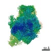 10985MC 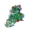 6yw5C 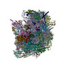 6yweC 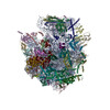 6ywsC 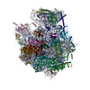 6ywvC  6ywxC M: map data used to model this data C: citing same article ( |
|---|---|
| Similar structure data |


-Links
 PDBj
PDBj


- Assembly
| Deposited unit | 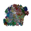
|
|---|---|
| 1 |
|
-Components
+RNA chain , 4 types, 4 molecules Aaabbee
+60S ribosomal protein ... , 3 types, 3 molecules BDe
+Related to ribosomal protein ... , 12 types, 12 molecules CEfKNUVWYaEEOO
+Protein , 44 types, 46 molecules FGgHLMPQRSX01235678i9bdAABBDDGGHHIIJJ...
+Ribosomal protein ... , 7 types, 7 molecules IJCCFFLLRRSS
+Mitochondrial ... , 3 types, 3 molecules ONNXX
+54S ribosomal protein ... , 2 types, 2 molecules Tc
+L51 S25 CI-B8 domain-containing ... , 2 types, 2 molecules 4j
+Probable ribosomal protein ... , 2 types, 2 molecules hMM
+37S ribosomal protein ... , 3 types, 3 molecules UUZZ11
+Protein/peptide , 1 types, 1 molecules cc
+Non-polymers , 6 types, 304 molecules 


+Details
-Experimental details
-Experiment
| Experiment | Method: ELECTRON MICROSCOPY |
|---|---|
| EM experiment | Aggregation state: PARTICLE / 3D reconstruction method: single particle reconstruction |
- Sample preparation
| Component | Name: Mitoribosome of Neurospora crassa in the tRNA bound state (P-site). Type: RIBOSOME / Entity ID: #1-#83 / Source: NATURAL | |||||||||||||||||||||||||||||||||||
|---|---|---|---|---|---|---|---|---|---|---|---|---|---|---|---|---|---|---|---|---|---|---|---|---|---|---|---|---|---|---|---|---|---|---|---|---|
| Molecular weight | Experimental value: NO | |||||||||||||||||||||||||||||||||||
| Source (natural) | Organism: Neurospora crassa (fungus) / Strain: K5-15-23-1 | |||||||||||||||||||||||||||||||||||
| Buffer solution | pH: 7.5 | |||||||||||||||||||||||||||||||||||
| Buffer component |
| |||||||||||||||||||||||||||||||||||
| Specimen | Embedding applied: NO / Shadowing applied: NO / Staining applied: NO / Vitrification applied: YES | |||||||||||||||||||||||||||||||||||
| Specimen support | Grid material: COPPER / Grid type: Quantifoil R2/2 | |||||||||||||||||||||||||||||||||||
| Vitrification | Instrument: FEI VITROBOT MARK IV / Cryogen name: ETHANE / Humidity: 100 % / Chamber temperature: 277 K |
- Electron microscopy imaging
| Experimental equipment |  Model: Titan Krios / Image courtesy: FEI Company |
|---|---|
| Microscopy | Model: FEI TITAN KRIOS |
| Electron gun | Electron source:  FIELD EMISSION GUN / Accelerating voltage: 300 kV / Illumination mode: FLOOD BEAM FIELD EMISSION GUN / Accelerating voltage: 300 kV / Illumination mode: FLOOD BEAM |
| Electron lens | Mode: BRIGHT FIELD / Calibrated magnification: 130000 X / C2 aperture diameter: 70 µm / Alignment procedure: COMA FREE |
| Specimen holder | Cryogen: NITROGEN / Specimen holder model: FEI TITAN KRIOS AUTOGRID HOLDER |
| Image recording | Electron dose: 35 e/Å2 / Detector mode: COUNTING / Film or detector model: GATAN K2 SUMMIT (4k x 4k) / Num. of real images: 3172 |
| EM imaging optics | Energyfilter name: GIF Quantum LS / Energyfilter slit width: 20 eV |
| Image scans | Movie frames/image: 20 / Used frames/image: 1-20 |
- Processing
| Software |
| ||||||||||||||||||||||||
|---|---|---|---|---|---|---|---|---|---|---|---|---|---|---|---|---|---|---|---|---|---|---|---|---|---|
| EM software |
| ||||||||||||||||||||||||
| CTF correction | Type: PHASE FLIPPING AND AMPLITUDE CORRECTION | ||||||||||||||||||||||||
| 3D reconstruction | Resolution: 3.05 Å / Resolution method: FSC 0.143 CUT-OFF / Num. of particles: 24601 / Symmetry type: POINT | ||||||||||||||||||||||||
| Refinement | Cross valid method: NONE Stereochemistry target values: GeoStd + Monomer Library + CDL v1.2 | ||||||||||||||||||||||||
| Displacement parameters | Biso mean: 21.41 Å2 | ||||||||||||||||||||||||
| Refine LS restraints |
|
