Movie
Movie Controller

[English] 日本語
 Yorodumi
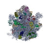
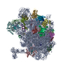
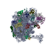
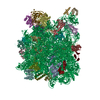
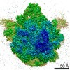
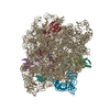
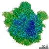
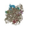
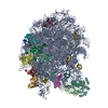
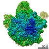
- PDB-6ys3: Cryo-EM structure of the 50S ribosomal subunit at 2.58 Angstroms ... -
+ Open data

- Basic information
| Entry | Database: PDB / ID: 6ys3 | ||||||
|---|---|---|---|---|---|---|---|
| Title | Cryo-EM structure of the 50S ribosomal subunit at 2.58 Angstroms with modeled GBC SecM peptide | ||||||
 Components Components |
| ||||||
 Keywords Keywords | RIBOSOME / translation 50S ribosome nascent peptide chain | ||||||
| Function / homology |  Function and homology information Function and homology informationstructural constituent of eye lens / lens development in camera-type eye / transcriptional attenuation / endoribonuclease inhibitor activity / positive regulation of ribosome biogenesis / RNA-binding transcription regulator activity / negative regulation of cytoplasmic translation / DnaA-L2 complex / translation repressor activity / visual perception ...structural constituent of eye lens / lens development in camera-type eye / transcriptional attenuation / endoribonuclease inhibitor activity / positive regulation of ribosome biogenesis / RNA-binding transcription regulator activity / negative regulation of cytoplasmic translation / DnaA-L2 complex / translation repressor activity / visual perception / negative regulation of DNA-templated DNA replication initiation / mRNA regulatory element binding translation repressor activity / response to reactive oxygen species / cytosolic ribosome assembly / ribosome assembly / assembly of large subunit precursor of preribosome / regulation of cell growth / DNA-templated transcription termination / response to radiation / mRNA 5'-UTR binding / large ribosomal subunit / transferase activity / ribosome binding / 5S rRNA binding / ribosomal large subunit assembly / large ribosomal subunit rRNA binding / cytosolic large ribosomal subunit / cytoplasmic translation / tRNA binding / negative regulation of translation / rRNA binding / structural constituent of ribosome / ribosome / translation / ribonucleoprotein complex / response to antibiotic / negative regulation of DNA-templated transcription / mRNA binding / DNA binding / RNA binding / zinc ion binding / cytosol / cytoplasm Similarity search - Function | ||||||
| Biological species |   | ||||||
| Method | ELECTRON MICROSCOPY / single particle reconstruction / cryo EM / Resolution: 2.58 Å | ||||||
 Authors Authors | Schulte, L. / Reitz, J. / Kudlinzki, D. / Hodirnau, V.V. / Frangakis, A. / Schwalbe, H. | ||||||
 Citation Citation | Journal: Nat Commun / Year: 2020 Title: Cysteine oxidation and disulfide formation in the ribosomal exit tunnel. Authors: Linda Schulte / Jiafei Mao / Julian Reitz / Sridhar Sreeramulu / Denis Kudlinzki / Victor-Valentin Hodirnau / Jakob Meier-Credo / Krishna Saxena / Florian Buhr / Julian D Langer / Martin ...Authors: Linda Schulte / Jiafei Mao / Julian Reitz / Sridhar Sreeramulu / Denis Kudlinzki / Victor-Valentin Hodirnau / Jakob Meier-Credo / Krishna Saxena / Florian Buhr / Julian D Langer / Martin Blackledge / Achilleas S Frangakis / Clemens Glaubitz / Harald Schwalbe /     Abstract: Understanding the conformational sampling of translation-arrested ribosome nascent chain complexes is key to understand co-translational folding. Up to now, coupling of cysteine oxidation, disulfide ...Understanding the conformational sampling of translation-arrested ribosome nascent chain complexes is key to understand co-translational folding. Up to now, coupling of cysteine oxidation, disulfide bond formation and structure formation in nascent chains has remained elusive. Here, we investigate the eye-lens protein γB-crystallin in the ribosomal exit tunnel. Using mass spectrometry, theoretical simulations, dynamic nuclear polarization-enhanced solid-state nuclear magnetic resonance and cryo-electron microscopy, we show that thiol groups of cysteine residues undergo S-glutathionylation and S-nitrosylation and form non-native disulfide bonds. Thus, covalent modification chemistry occurs already prior to nascent chain release as the ribosome exit tunnel provides sufficient space even for disulfide bond formation which can guide protein folding. | ||||||
| History |
|
- Structure visualization
| Movie |
Movie viewer |
|---|---|
| Structure viewer | Molecule: MolmilJmol/JSmol |
- Downloads & links
-Download
| PDBx/mmCIF format | 6ys3.cif.gz | 2 MB | Display | PDBx/mmCIF format |
|---|---|---|---|---|
| PDB format | pdb6ys3.ent.gz | 1.5 MB | Display | PDB format |
| PDBx/mmJSON format | 6ys3.json.gz | Tree view | PDBx/mmJSON format | |
| Others |  Other downloads Other downloads |
-Validation report
| Arichive directory | https://data.pdbj.org/pub/pdb/validation_reports/ys/6ys3ftp://data.pdbj.org/pub/pdb/validation_reports/ys/6ys3 | HTTPS FTP |
|---|
-Related structure data
| Related structure data | 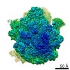 10891MC 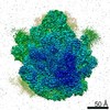 4531C  6qdwC M: map data used to model this data C: citing same article ( |
|---|---|
| Similar structure data |

-Links
 PDBj
PDBj


- Assembly
| Deposited unit | 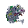
|
|---|---|
| 1 |
|
-Components
+50S ribosomal protein ... , 28 types, 28 molecules 01234678cdefghjklmnopqrstuwy
-RNA chain , 3 types, 3 molecules abv
| #9: RNA chain | Mass: 38790.090 Da / Num. of mol.: 1 / Source method: isolated from a natural source / Source: (natural) |
|---|---|
| #10: RNA chain | Mass: 942342.938 Da / Num. of mol.: 1 / Source method: isolated from a natural source / Source: (natural) |
| #29: RNA chain | Mass: 23935.143 Da / Num. of mol.: 1 / Source method: isolated from a natural source / Source: (natural) |
-Protein , 1 types, 1 molecules z
| #32: Protein | Mass: 7157.970 Da / Num. of mol.: 1 Source method: isolated from a genetically manipulated source Source: (gene. exp.) |
|---|
-Details
| Has protein modification | Y |
|---|
-Experimental details
-Experiment
| Experiment | Method: ELECTRON MICROSCOPY |
|---|---|
| EM experiment | Aggregation state: PARTICLE / 3D reconstruction method: single particle reconstruction |
- Sample preparation
| Component |
| |||||||||||||||||||||||||
|---|---|---|---|---|---|---|---|---|---|---|---|---|---|---|---|---|---|---|---|---|---|---|---|---|---|---|
| Source (natural) |
| |||||||||||||||||||||||||
| Source (recombinant) | Organism: | |||||||||||||||||||||||||
| Buffer solution | pH: 7.5 | |||||||||||||||||||||||||
| Buffer component |
| |||||||||||||||||||||||||
| Specimen | Conc.: 0.12 mg/ml / Embedding applied: NO / Shadowing applied: NO / Staining applied: NO / Vitrification applied: YES | |||||||||||||||||||||||||
| Specimen support | Grid material: COPPER / Grid mesh size: 300 divisions/in. / Grid type: Quantifoil R2/1 | |||||||||||||||||||||||||
| Vitrification | Instrument: FEI VITROBOT MARK IV / Cryogen name: ETHANE / Humidity: 100 % / Chamber temperature: 277.15 K |
- Electron microscopy imaging
| Experimental equipment |  Model: Titan Krios / Image courtesy: FEI Company |
|---|---|
| Microscopy | Model: FEI TITAN KRIOS |
| Electron gun | Electron source:  FIELD EMISSION GUN / Accelerating voltage: 300 kV / Illumination mode: FLOOD BEAM FIELD EMISSION GUN / Accelerating voltage: 300 kV / Illumination mode: FLOOD BEAM |
| Electron lens | Mode: BRIGHT FIELD / Cs: 2.7 mm |
| Specimen holder | Cryogen: NITROGEN / Specimen holder model: FEI TITAN KRIOS AUTOGRID HOLDER |
| Image recording | Average exposure time: 7.2 sec. / Electron dose: 60 e/Å2 / Detector mode: COUNTING / Film or detector model: GATAN K2 SUMMIT (4k x 4k) |
| Image scans | Movie frames/image: 36 |
- Processing
| EM software |
| ||||||||||||||||||||
|---|---|---|---|---|---|---|---|---|---|---|---|---|---|---|---|---|---|---|---|---|---|
| CTF correction | Type: PHASE FLIPPING AND AMPLITUDE CORRECTION | ||||||||||||||||||||
| Particle selection | Num. of particles selected: 436999 | ||||||||||||||||||||
| Symmetry | Point symmetry: C1 (asymmetric) | ||||||||||||||||||||
| 3D reconstruction | Resolution: 2.58 Å / Resolution method: FSC 0.143 CUT-OFF / Num. of particles: 196254 / Symmetry type: POINT | ||||||||||||||||||||
| Atomic model building | Protocol: RIGID BODY FIT / Space: REAL | ||||||||||||||||||||
| Atomic model building | PDB-ID: 3JBU Accession code: 3JBU / Source name: PDB / Type: experimental model |