stringent response / translational termination / transcriptional attenuation / endoribonuclease inhibitor activity / positive regulation of ribosome biogenesis / RNA-binding transcription regulator activity / negative regulation of cytoplasmic translation / DnaA-L2 complex / translation repressor activity / negative regulation of DNA-templated DNA replication initiation ...stringent response / translational termination / transcriptional attenuation / endoribonuclease inhibitor activity / positive regulation of ribosome biogenesis / RNA-binding transcription regulator activity / negative regulation of cytoplasmic translation / DnaA-L2 complex / translation repressor activity / negative regulation of DNA-templated DNA replication initiation / mRNA regulatory element binding translation repressor activity / response to reactive oxygen species / cytosolic ribosome assembly / ribosome assembly / assembly of large subunit precursor of preribosome / regulation of cell growth / DNA-templated transcription termination / response to radiation / mRNA 5'-UTR binding / large ribosomal subunit / transferase activity / ribosome binding / 5S rRNA binding / ribosomal large subunit assembly / large ribosomal subunit rRNA binding / cytosolic large ribosomal subunit / cytoplasmic translation / tRNA binding / negative regulation of translation / rRNA binding / structural constituent of ribosome / ribosome / translation / response to antibiotic / negative regulation of DNA-templated transcription / mRNA binding / DNA binding / RNA binding / zinc ion binding / cytoplasm / cytosol Similarity search - Function
Ribosomal protein L10, eubacterial, conserved site / Ribosomal protein L10 signature. / Ribosomal protein L10 / : / Ribosomal protein L11, bacterial-type / Ribosomal protein L25, short-form / Ribosomal protein L11, conserved site / Ribosomal protein L11 signature. / Ribosomal protein L10-like domain superfamily / Ribosomal protein L10P ...Ribosomal protein L10, eubacterial, conserved site / Ribosomal protein L10 signature. / Ribosomal protein L10 / : / Ribosomal protein L11, bacterial-type / Ribosomal protein L25, short-form / Ribosomal protein L11, conserved site / Ribosomal protein L11 signature. / Ribosomal protein L10-like domain superfamily / Ribosomal protein L10P / Ribosomal protein L10 / Ribosomal protein L9 signature. / Ribosomal protein L16 signature 1. / Ribosomal protein L6, conserved site / Ribosomal protein L6 signature 1. / Ribosomal protein L9, bacteria/chloroplast / Ribosomal protein L9, C-terminal / Ribosomal protein L9, C-terminal domain / Ribosomal protein L21, conserved site / Ribosomal protein L21 signature. / : / Ribosomal protein L11, N-terminal / Ribosomal protein L9, C-terminal domain superfamily / Ribosomal protein L11, N-terminal domain / Ribosomal protein L11/L12 / Ribosomal protein L11, C-terminal / Ribosomal protein L11, C-terminal domain superfamily / Ribosomal protein L11/L12, N-terminal domain superfamily / Ribosomal protein L11/L12 / Ribosomal protein L11, RNA binding domain / Ribosomal protein L16 signature 2. / Ribosomal protein L16, conserved site / Ribosomal protein L17 signature. / Ribosomal L25p family / Ribosomal protein L25 / Ribosomal protein L36 signature. / Ribosomal protein L25/Gln-tRNA synthetase, N-terminal / Ribosomal protein L25/Gln-tRNA synthetase, anti-codon-binding domain superfamily / : / Ribosomal protein L28/L24 superfamily / Ribosomal protein L33, conserved site / Ribosomal protein L33 signature. / Ribosomal protein L32p, bacterial type / Ribosomal protein L35, conserved site / Ribosomal protein L35 signature. / Ribosomal protein L9 / Ribosomal protein L9, N-terminal domain superfamily / Ribosomal protein L9, N-terminal / Ribosomal protein L9, N-terminal domain / Ribosomal protein L28 / Ribosomal protein L35, non-mitochondrial / Ribosomal protein L18, bacterial-type / : / Ribosomal protein L6, bacterial-type / Ribosomal protein L5, bacterial-type / Ribosomal protein L9/RNase H1, N-terminal / Ribosomal protein L19, conserved site / Ribosomal protein L19 signature. / : / Ribosomal protein L36 / Ribosomal protein L36 superfamily / Ribosomal protein L36 / Ribosomal protein L20 signature. / Ribosomal protein L34, conserved site / Ribosomal protein L34 signature. / Ribosomal protein L14P, bacterial-type / Ribosomal protein L27, conserved site / Ribosomal protein L27 signature. / Ribosomal protein L35 / Ribosomal protein L35 superfamily / Ribosomal protein L22, bacterial/chloroplast-type / Ribosomal protein L35 / Ribosomal protein L33 / Ribosomal protein L18 / Ribosomal L18 of archaea, bacteria, mitoch. and chloroplast / Ribosomal protein L33 / Ribosomal protein L2, bacterial/organellar-type / Ribosomal L28 family / Ribosomal protein L33 superfamily / Ribosomal protein L28/L24 / Ribosomal protein L30, bacterial-type / L28p-like / Ribosomal protein L16 / Ribosomal protein L20 / Ribosomal protein L20 / Ribosomal protein L20, C-terminal / Ribosomal protein L19 / Ribosomal protein L19 / Ribosomal protein L19 superfamily / : / Large ribosomal subunit protein uL24, C-terminal domain / Ribosomal protein L17 / Ribosomal protein L17 superfamily / Ribosomal protein L17 / Ribosomal protein L27 / Ribosomal L27 protein / Ribosomal protein L34 / Ribosomal protein L34 / Ribosomal protein L24 / Ribosomal L32p protein family Similarity search - Domain/homology
Large ribosomal subunit protein uL15 / Large ribosomal subunit protein uL10 / Large ribosomal subunit protein uL11 / Large ribosomal subunit protein bL19 / Large ribosomal subunit protein bL20 / Large ribosomal subunit protein bL27 / Large ribosomal subunit protein bL28 / Large ribosomal subunit protein uL29 / Large ribosomal subunit protein bL32 / Large ribosomal subunit protein bL33 ...Large ribosomal subunit protein uL15 / Large ribosomal subunit protein uL10 / Large ribosomal subunit protein uL11 / Large ribosomal subunit protein bL19 / Large ribosomal subunit protein bL20 / Large ribosomal subunit protein bL27 / Large ribosomal subunit protein bL28 / Large ribosomal subunit protein uL29 / Large ribosomal subunit protein bL32 / Large ribosomal subunit protein bL33 / Large ribosomal subunit protein bL34 / Large ribosomal subunit protein bL35 / Large ribosomal subunit protein bL36A / Large ribosomal subunit protein bL9 / Large ribosomal subunit protein uL13 / Large ribosomal subunit protein uL14 / Large ribosomal subunit protein uL16 / Large ribosomal subunit protein uL23 / Large ribosomal subunit protein bL17 / Large ribosomal subunit protein bL21 / Large ribosomal subunit protein uL30 / Large ribosomal subunit protein uL6 / Large ribosomal subunit protein uL18 / Large ribosomal subunit protein uL2 / Large ribosomal subunit protein uL3 / Large ribosomal subunit protein uL24 / Large ribosomal subunit protein uL4 / Large ribosomal subunit protein uL22 / Large ribosomal subunit protein uL5 / Large ribosomal subunit protein bL25 Similarity search - Component
Biological species
Escherichia coli (E. coli) / Escherichia coli (strain K12) (bacteria)
Method
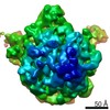
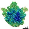
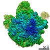
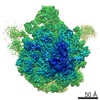
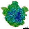
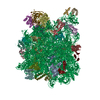
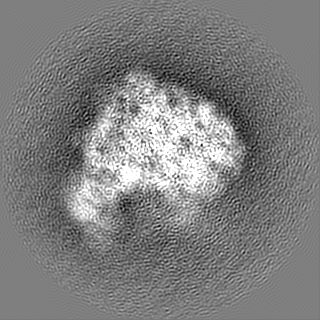
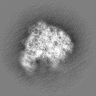
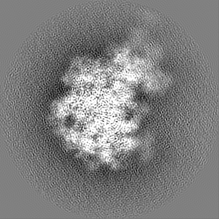
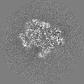
single particle reconstruction / cryo EM / Resolution: 3.14 Å
National Natural Science Foundation of China (NSFC)
31630087
China
Ministry of Science and Technology (MoST, China)
2016YFA0500700
China
Other government
China Postdoctoral Science Foundation 1131000065
China
Other government
Shenzhen Science and Technology Innovation Committee (JCYJ20180302174213122)
China
Citation
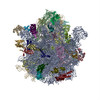
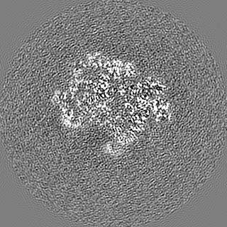
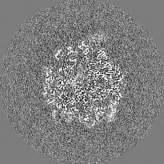
Journal: Proc Natl Acad Sci U S A / Year: 2020 Title: Loss of a single methylation in 23S rRNA delays 50S assembly at multiple late stages and impairs translation initiation and elongation. Authors: Wei Wang / Wanqiu Li / Xueliang Ge / Kaige Yan / Chandra Sekhar Mandava / Suparna Sanyal / Ning Gao / Abstract: Ribosome biogenesis is a complex process, and dozens of factors are required to facilitate and regulate the subunit assembly in bacteria. The 2'-O-methylation of U2552 in 23S rRNA by ...Ribosome biogenesis is a complex process, and dozens of factors are required to facilitate and regulate the subunit assembly in bacteria. The 2'-O-methylation of U2552 in 23S rRNA by methyltransferase RrmJ is a crucial step in late-stage assembly of the 50S subunit. Its absence results in severe growth defect and marked accumulation of pre50S assembly intermediates. In the present work, we employed cryoelectron microscopy to characterize a set of late-stage pre50S particles isolated from an Δ strain. These assembly intermediates (solved at 3.2 to 3.8 Å resolution) define a collection of late-stage particles on a progressive assembly pathway. Apart from the absence of L16, L35, and L36, major structural differences between these intermediates and the mature 50S subunit are clustered near the peptidyl transferase center, such as H38, H68-71, and H89-93. In addition, the ribosomal A-loop of the mature 50S subunit from Δ strain displays large local flexibility on nucleotides next to unmethylated U2552. Fast kinetics-based biochemical assays demonstrate that the Δ 50S subunit is only 50% active and two times slower than the WT 50S subunit in rapid subunit association. While the Δ 70S ribosomes show no defect in peptide bond formation, peptide release, and ribosome recycling, they translocate with 20% slower rate than the WT ribosomes in each round of elongation. These defects amplify during synthesis of the full-length proteins and cause overall defect in protein synthesis. In conclusion, our data reveal the molecular roles of U2552 methylation in both ribosome biogenesis and protein translation.
History
Deposition
Apr 9, 2020
-
Header (metadata) release
Jul 1, 2020
-
Map release
Jul 1, 2020
-
Update
Mar 27, 2024
-
Current status
Mar 27, 2024
Processing site: PDBj / Status: Released
-
Structure visualization
Movie
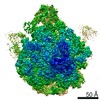
Surface view with section colored by density value
pH: 7.5 Details: Solutions were made fresh form concentrated to avoid microbial contamination.
Grid
Model: Quantifoil R2/2 / Material: COPPER / Mesh: 300 / Support film - Material: CARBON / Support film - topology: CONTINUOUS / Support film - Film thickness: 7000 / Pretreatment - Type: GLOW DISCHARGE / Pretreatment - Time: 30 sec. / Pretreatment - Atmosphere: AIR / Pretreatment - Pressure: 0.03 kPa / Details: The grid was coated with continuous carbon film.
Vitrification
Cryogen name: ETHANE / Chamber humidity: 100 % / Chamber temperature: 277 K / Instrument: FEI VITROBOT MARK IV / Details: blot for 2 seconds before plunging.
Details
This sample was monodisperse.
-
Electron microscopy
Microscope
FEI TITAN KRIOS
Details
Preliminary grid screening was performed manually.
Image recording
Film or detector model: FEI FALCON II (4k x 4k) / Digitization - Dimensions - Width: 4096 pixel / Digitization - Dimensions - Height: 4096 pixel / Digitization - Frames/image: 3-28 / Number grids imaged: 1 / Number real images: 1164 / Average electron dose: 45.0 e/Å2
Electron beam
Acceleration voltage: 300 kV / Electron source: FIELD EMISSION GUN
Number selected: 233762 Details: Images were binned and lowpass filtered for autopicking.
Startup model
Type of model: OTHER Details: The model was a low resolution 50S ribosome generated from our own data.
Final reconstruction
Number classes used: 1 / Applied symmetry - Point group: C1 (asymmetric) / Algorithm: SIMULTANEOUS ITERATIVE (SIRT) / Resolution.type: BY AUTHOR / Resolution: 3.14 Å / Resolution method: FSC 0.143 CUT-OFF / Software - Name: RELION (ver. 1.4) Details: A soft mask was added to improve resolution at the final refinement. Number images used: 98194
Initial angle assignment
Type: MAXIMUM LIKELIHOOD / Software - Name: RELION (ver. 1.4) / Details: Relion software was used in the reconstruction.
Final angle assignment
Type: MAXIMUM LIKELIHOOD / Software - Name: RELION (ver. 1.4) / Details: Relion software was used in the reconstruction.
In the structure databanks used in Yorodumi, some data are registered as the other names, "COVID-19 virus" and "2019-nCoV". Here are the details of the virus and the list of structure data.
Jan 31, 2019. EMDB accession codes are about to change! (news from PDBe EMDB page)
EMDB accession codes are about to change! (news from PDBe EMDB page)
The allocation of 4 digits for EMDB accession codes will soon come to an end. Whilst these codes will remain in use, new EMDB accession codes will include an additional digit and will expand incrementally as the available range of codes is exhausted. The current 4-digit format prefixed with “EMD-” (i.e. EMD-XXXX) will advance to a 5-digit format (i.e. EMD-XXXXX), and so on. It is currently estimated that the 4-digit codes will be depleted around Spring 2019, at which point the 5-digit format will come into force.
The EM Navigator/Yorodumi systems omit the EMD- prefix.
Related info.:Q: What is EMD? / ID/Accession-code notation in Yorodumi/EM Navigator
Yorodumi is a browser for structure data from EMDB, PDB, SASBDB, etc.
This page is also the successor to EM Navigator detail page, and also detail information page/front-end page for Omokage search.
The word "yorodu" (or yorozu) is an old Japanese word meaning "ten thousand". "mi" (miru) is to see.
Related info.:EMDB / PDB / SASBDB / Comparison of 3 databanks / Yorodumi Search / Aug 31, 2016. New EM Navigator & Yorodumi / Yorodumi Papers / Jmol/JSmol / Function and homology information / Changes in new EM Navigator and Yorodumi
 Movie
Movie Controller


 Open data
 Basic information
 Map data
 Sample
 Keywords
 Function and homology information
 Authors
 China, 4 items
  Citation

 Structure visualization
 Downloads & links
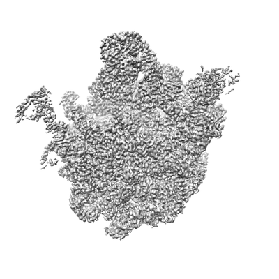 emd_30215.png
 http://ftp.pdbj.org/pub/emdb/structures/EMD-30215
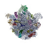


 Z (Sec.)
 Y (Row.)
 X (Col.)


 Sample components
 Processing
 Electron microscopy
 FIELD EMISSION GUN