National Institutes of Health/National Institute on Aging (NIH/NIA)
RF1AG065341
United States
National Institutes of Health/National Institute on Aging (NIH/NIA)
P30AG072979
United States
National Institutes of Health/National Institute on Aging (NIH/NIA)
P01AG066597
United States
National Institutes of Health/National Institute on Aging (NIH/NIA)
U19AG062418
United States
David and Lucile Packard Foundation
2019-69645
United States
Burroughs Wellcome Fund
1022785
United States
National Institutes of Health/National Institute of General Medical Sciences (NIH/NIGMS)
RM1GM136511
United States
National Institutes of Health/National Institute on Aging (NIH/NIA)
F30AG077756
United States
National Institutes of Health/National Cancer Institute (NIH/NCI)
F30CA261198
United States
National Institutes of Health/National Institute of General Medical Sciences (NIH/NIGMS)
R35GM130302
United States
National Institutes of Health/National Institute of General Medical Sciences (NIH/NIGMS)
T32GM132039
United States
Citation
Journal: Nat Commun / Year: 2024 Title: Ultrastructure of human brain tissue vitrified from autopsy revealed by cryo-ET with cryo-plasma FIB milling. Authors: Benjamin C Creekmore / Kathryn Kixmoeller / Ben E Black / Edward B Lee / Yi-Wei Chang / Abstract: Ultrastructure of human brain tissue has traditionally been examined using electron microscopy (EM) following fixation, staining, and sectioning, which limit resolution and introduce artifacts. ...Ultrastructure of human brain tissue has traditionally been examined using electron microscopy (EM) following fixation, staining, and sectioning, which limit resolution and introduce artifacts. Alternatively, cryo-electron tomography (cryo-ET) allows higher resolution imaging of unfixed cellular samples while preserving architecture, but it requires samples to be vitreous and thin enough for transmission EM. Due to these requirements, cryo-ET has yet to be employed to investigate unfixed, never previously frozen human brain tissue. Here we present a method for generating lamellae in human brain tissue obtained at time of autopsy that can be imaged via cryo-ET. We vitrify the tissue via plunge-freezing and use xenon plasma focused ion beam (FIB) milling to generate lamellae directly on-grid at variable depth inside the tissue. Lamellae generated in Alzheimer's disease brain tissue reveal intact subcellular structures including components of autophagy and potential pathologic tau fibrils. Furthermore, we reveal intact compact myelin and functional cytoplasmic expansions. These images indicate that plasma FIB milling with cryo-ET may be used to elucidate nanoscale structures within the human brain.
Cryogen name: ETHANE-PROPANE / Chamber humidity: 90 % / Chamber temperature: 288 K / Instrument: LEICA EM GP / Details: back blot for 6s before plunging.
Cryo protectant
20% glycerol, 1M trehalose
Sectioning
Focused ion beam - Instrument: OTHER / Focused ion beam - Ion: OTHER / Focused ion beam - Voltage: 30 / Focused ion beam - Current: 0.05 / Focused ion beam - Duration: 7200 / Focused ion beam - Temperature: 113 K / Focused ion beam - Initial thickness: 100000 / Focused ion beam - Final thickness: 350 Focused ion beam - Details: The value given for _em_focused_ion_beam.instrument is Tescan S8000X. This is not in a list of allowed values {'DB235', 'OTHER'} so OTHER is written into the XML file.
-
Electron microscopy
Microscope
FEI TITAN KRIOS
Specialist optics
Energy filter - Name: GIF Bioquantum / Energy filter - Slit width: 20 eV
Image recording
Film or detector model: GATAN K3 (6k x 4k) / Average electron dose: 3.4 e/Å2
Electron beam
Acceleration voltage: 300 kV / Electron source: FIELD EMISSION GUN
Raw images were gain normalize in serialEM. Raw images were high-pass filtered at 1000 pixels then vertical line filtered with a 95% tolerance of direction.
Final reconstruction
Algorithm: BACK PROJECTION / Number images used: 25
+
About Yorodumi
-
News
-
Feb 9, 2022. New format data for meta-information of EMDB entries
New format data for meta-information of EMDB entries
Version 3 of the EMDB header file is now the official format.
The previous official version 1.9 will be removed from the archive.
In the structure databanks used in Yorodumi, some data are registered as the other names, "COVID-19 virus" and "2019-nCoV". Here are the details of the virus and the list of structure data.
Jan 31, 2019. EMDB accession codes are about to change! (news from PDBe EMDB page)
EMDB accession codes are about to change! (news from PDBe EMDB page)
The allocation of 4 digits for EMDB accession codes will soon come to an end. Whilst these codes will remain in use, new EMDB accession codes will include an additional digit and will expand incrementally as the available range of codes is exhausted. The current 4-digit format prefixed with “EMD-” (i.e. EMD-XXXX) will advance to a 5-digit format (i.e. EMD-XXXXX), and so on. It is currently estimated that the 4-digit codes will be depleted around Spring 2019, at which point the 5-digit format will come into force.
The EM Navigator/Yorodumi systems omit the EMD- prefix.
Related info.:Q: What is EMD? / ID/Accession-code notation in Yorodumi/EM Navigator
Yorodumi is a browser for structure data from EMDB, PDB, SASBDB, etc.
This page is also the successor to EM Navigator detail page, and also detail information page/front-end page for Omokage search.
The word "yorodu" (or yorozu) is an old Japanese word meaning "ten thousand". "mi" (miru) is to see.
Related info.:EMDB / PDB / SASBDB / Comparison of 3 databanks / Yorodumi Search / Aug 31, 2016. New EM Navigator & Yorodumi / Yorodumi Papers / Jmol/JSmol / Function and homology information / Changes in new EM Navigator and Yorodumi
 Movie
Movie Controller

 Yorodumi
 Open data
 Basic information
 Map data
 Sample
 Keywords
 Homo sapiens (human)
 Authors
 United States, 13 items
  Citation
 Structure visualization
 Downloads & links
 EMDB map data format
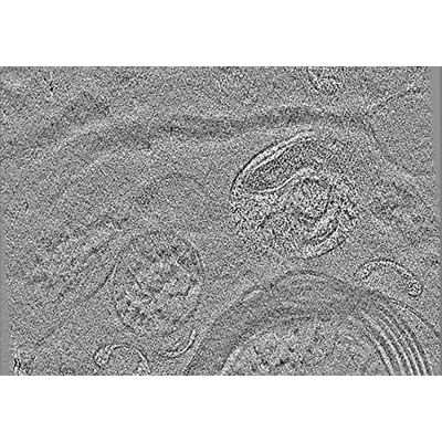 emd_43330.png
 http://ftp.pdbj.org/pub/emdb/structures/EMD-43330

 Z (Sec.)
 Y (Row.)
 X (Col.)


 Sample components
 Processing
 Electron microscopy
 FIELD EMISSION GUN
