 ムービー
ムービー コントローラー
 構造ビューア
 EMN検索について


-検索条件
-検索結果
検索 (著者・登録者: bennet & p)の結果67件中、1から50件目までを表示しています
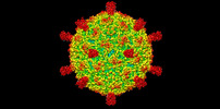
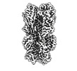
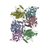
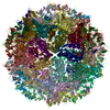
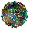
EMDB-45583: 
The Structure of Spiroplasma Virus 4
PDB-9cgm:
The Structure of Spiroplasma Virus 4

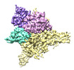
EMDB-40853:
CH505 Disulfide Stapled SOSIP Bound to b12 Fab

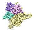
EMDB-40854:
CH505 Disulfide Stapled SOSIP Bound to CH235.12 Fab
PDB-8sxi:
CH505 Disulfide Stapled SOSIP Bound to b12 Fab

PDB-8sxj:
CH505 Disulfide Stapled SOSIP Bound to CH235.12 Fab

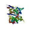
EMDB-16986:
Structure of the relaxed thin filament from FIB milled left ventricular mouse myofibrils (tropomyosin masked out)

EMDB-16987:
Structure of the relaxed thin filament from FIB milled left ventricular mouse myofibrils (including tropomyosin)

EMDB-16988:
Tomogram of sarcomere C-zone from mouse cardiac muscle

EMDB-16989:
Tomogram of sarcomere M-band to C-zone from mouse cardiac muscle

EMDB-16990:
Structure of the relaxed thick filament from FIB milled left ventricular mouse myofibrils - Crowns P2-A1

EMDB-16991:
Structure of the relaxed thick filament from FIB milled left ventricular mouse myofibrils - M-band

EMDB-16992:
Structure of the relaxed thick filament from FIB milled left ventricular mouse myofibrils - Crowns A15-A29

EMDB-16993:
Structure of the relaxed thick filament from FIB milled left ventricular mouse myofibrils - Crown P1

EMDB-16994:
Structure of the relaxed thick filament from FIB milled left ventricular mouse myofibrils - Crowns A11-A15

EMDB-16995:
Structure of the relaxed thick filament from FIB milled left ventricular mouse myofibrils - Crowns A8-A12

EMDB-16996:
Structure of the relaxed thick filament from FIB milled left ventricular mouse myofibrils - Crowns A5-A7

EMDB-16997:
Structure of the relaxed thick filament from FIB milled left ventricular mouse myofibrils - Crowns A1-A5

EMDB-18146:
In situ structures from relaxed cardiac myofibrils reveal the organization of the muscle thick filament

EMDB-18200:
Thin filament consensus map from FIB milled relaxed left ventricular mouse myofibrils

EMDB-18147:
Thin filament from FIB milled relaxed left ventricular mouse myofibrils

EMDB-18198:
Helical reconstruction of the relaxed thick filament from FIB milled left ventricular mouse myofibrils
PDB-8q4g:
Thin filament from FIB milled relaxed left ventricular mouse myofibrils

PDB-8q6t:
Helical reconstruction of the relaxed thick filament from FIB milled left ventricular mouse myofibrils

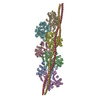
EMDB-26977:
CryoEM structure of human S-OPA1 assembled on lipid membrane in membrane-adjacent state

EMDB-26984:
CryoEM structure of human S-OPA1 assembled on lipid membrane in membrane-distal state

PDB-8ct1:
CryoEM structure of human S-OPA1 assembled on lipid membrane in membrane-adjacent state

PDB-8ct9:
CryoEM structure of human S-OPA1 assembled on lipid membrane in membrane-distal state
EMDB-40557:
Cryo-EM structure of designed Influenza HA binder, HA_20, bound to Influenza HA (Strain: Iowa43)

PDB-8sk7:
Cryo-EM structure of designed Influenza HA binder, HA_20, bound to Influenza HA (Strain: Iowa43)

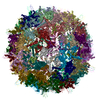
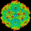
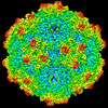
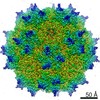
EMDB-29377:
AAV1 VP3 Only Capsid

PDB-8fq4:
AAV1 VP3 Only Capsid
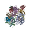
EMDB-27898:
Cryo-EM structure of human glycerol-3-phosphate acyltransferase 1 (GPAT1) in complex with 2-oxohexadecyl-CoA
EMDB-27899:
Cryo-EM structure of human glycerol-3-phosphate acyltransferase 1 (GPAT1) in complex with CoA and palmitoyl-LPA
PDB-8e4y:
Cryo-EM structure of human glycerol-3-phosphate acyltransferase 1 (GPAT1) in complex with 2-oxohexadecyl-CoA

PDB-8e50:
Cryo-EM structure of human glycerol-3-phosphate acyltransferase 1 (GPAT1) in complex with CoA and palmitoyl-LPA

EMDB-26595:
CryoEM structure of human LACTB filament
PDB-7ulw:
CryoEM structure of human LACTB filament
EMDB-26390:
SAAV pH 7.4 capsid structure
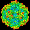
EMDB-26391:
SAAV pH 6.0 capsid structure
EMDB-26392:
SAAV pH 5.5 capsid structure
EMDB-26393:
SAAV pH 4.0 capsid structure
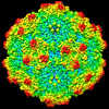
EMDB-26394:
The SAAV capsid in complex with 3'SLN
EMDB-26395:
The SAAV capsid in complex with 6'SLN
PDB-7u94:
SAAV pH 7.4 capsid structure

PDB-7u95:
SAAV pH 6.0 capsid structure
PDB-7u96:
SAAV pH 5.5 capsid structure

PDB-7u97:
SAAV pH 4.0 capsid structure
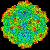
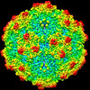
EMDB-24266:
Cryo-EM structure of AAV True Type

PDB-7na6:
Cryo-EM structure of AAV True Type
ページ:
 wwPDBはEMDBデータモデルのバージョン3へ移行します
