 Movie
Movie Controller

[English] 日本語
 Yorodumi
- EMDB-27899: Cryo-EM structure of human glycerol-3-phosphate acyltransferase 1... -
+ Open data

- Basic information
| Entry |  | |||||||||
|---|---|---|---|---|---|---|---|---|---|---|
| Title | Cryo-EM structure of human glycerol-3-phosphate acyltransferase 1 (GPAT1) in complex with CoA and palmitoyl-LPA | |||||||||
 Map data Map data | Half map 1 | |||||||||
 Sample Sample |
| |||||||||
 Keywords Keywords | acyltransferase / LPA / monotopic / mitochondrial / membrane protein | |||||||||
| Function / homology |  Function and homology information Function and homology informationglycerol-3-phosphate 1-O-acyltransferase / glycerol-3-phosphate O-acyltransferase activity / phosphatidylglycerol biosynthetic process / CDP-diacylglycerol biosynthetic process / Triglyceride biosynthesis / diacylglycerol biosynthetic process / glycerophospholipid metabolic process / negative regulation of activation-induced cell death of T cells / phosphatidic acid biosynthetic process / Synthesis of PA ...glycerol-3-phosphate 1-O-acyltransferase / glycerol-3-phosphate O-acyltransferase activity / phosphatidylglycerol biosynthetic process / CDP-diacylglycerol biosynthetic process / Triglyceride biosynthesis / diacylglycerol biosynthetic process / glycerophospholipid metabolic process / negative regulation of activation-induced cell death of T cells / phosphatidic acid biosynthetic process / Synthesis of PA / acyl-CoA metabolic process / activation-induced cell death of T cells / triglyceride biosynthetic process / phospholipid homeostasis / activated T cell proliferation / positive regulation of multicellular organism growth / positive regulation of activated T cell proliferation / RUNX1 regulates estrogen receptor mediated transcription / fatty acid homeostasis / response to glucose / Activation of gene expression by SREBF (SREBP) / regulation of cytokine production / fatty acid metabolic process / MLL4 and MLL3 complexes regulate expression of PPARG target genes in adipogenesis and hepatic steatosis / Estrogen-dependent gene expression / defense response to virus / mitochondrial outer membrane / mitochondrion / plasma membrane Similarity search - Function | |||||||||
| Biological species |  Homo sapiens (human) Homo sapiens (human) | |||||||||
| Method | single particle reconstruction / cryo EM / Resolution: 3.67 Å | |||||||||
 Authors Authors | Wasilko DJ / Johnson ZL / Ammirati M / Chang JS / Han S / Wu H | |||||||||
| Funding support | 1 items
| |||||||||
 Citation Citation | Journal: Nat Struct Mol Biol / Year: 2023 Title: Structural basis of the acyl-transfer mechanism of human GPAT1. Authors: Zachary Lee Johnson / Mark Ammirati / David Jonathan Wasilko / Jeanne S Chang / Stephen Noell / Timothy L Foley / Hyejin Yoon / Kathleen Smith / Shoh Asano / Katherine Hales / Min Wan / ...Authors: Zachary Lee Johnson / Mark Ammirati / David Jonathan Wasilko / Jeanne S Chang / Stephen Noell / Timothy L Foley / Hyejin Yoon / Kathleen Smith / Shoh Asano / Katherine Hales / Min Wan / Qingyi Yang / Mary A Piotrowski / Kathleen A Farley / Tamara Gilbert / Lisa M Aschenbrenner / Kimberly F Fennell / Jason K Dutra / Mary Xu / Chunyang Guo / Alison E Varghese / Justin Bellenger / Alandra Quinn / Christopher W Am Ende / Graham M West / Matthew C Griffor / Donald Bennett / Matthew Calabrese / Claire M Steppan / Seungil Han / Huixian Wu /  Abstract: Glycerol-3-phosphate acyltransferase (GPAT)1 is a mitochondrial outer membrane protein that catalyzes the first step of de novo glycerolipid biosynthesis. Hepatic expression of GPAT1 is linked to ...Glycerol-3-phosphate acyltransferase (GPAT)1 is a mitochondrial outer membrane protein that catalyzes the first step of de novo glycerolipid biosynthesis. Hepatic expression of GPAT1 is linked to liver fat accumulation and the severity of nonalcoholic fatty liver diseases. Here we present the cryo-EM structures of human GPAT1 in substrate analog-bound and product-bound states. The structures reveal an N-terminal acyltransferase domain that harbors important catalytic motifs and a tightly associated C-terminal domain that is critical for proper protein folding. Unexpectedly, GPAT1 has no transmembrane regions as previously proposed but instead associates with the membrane via an amphipathic surface patch and an N-terminal loop-helix region that contains a mitochondrial-targeting signal. Combined structural, computational and functional studies uncover a hydrophobic pathway within GPAT1 for lipid trafficking. The results presented herein lay a framework for rational inhibitor development for GPAT1. | |||||||||
| History |
|
- Structure visualization
| Supplemental images |
|---|

- Downloads & links
-EMDB archive
| Map data | emd_27899.map.gz | 202.4 MB | EMDB map data format | |
|---|---|---|---|---|
| Header (meta data) | emd-27899-v30.xmlemd-27899.xml | 21.6 KB 21.6 KB | Display Display | EMDB header |
| FSC (resolution estimation) | emd_27899_fsc.xml | 13.7 KB | Display | FSC data file |
| Images | 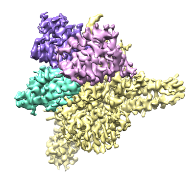 emd_27899.png emd_27899.png | 137.2 KB | ||
| Filedesc metadata | emd-27899.cif.gz | 7.1 KB | ||
| Others | emd_27899_half_map_1.map.gzemd_27899_half_map_2.map.gz | 171.6 MB 171 MB | ||
| Archive directory |  http://ftp.pdbj.org/pub/emdb/structures/EMD-27899ftp://ftp.pdbj.org/pub/emdb/structures/EMD-27899 http://ftp.pdbj.org/pub/emdb/structures/EMD-27899ftp://ftp.pdbj.org/pub/emdb/structures/EMD-27899 | HTTPS FTP |
-Related structure data
| Related structure data |  8e50MC 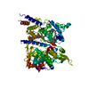 8e4yC C: citing same article ( M: atomic model generated by this map |
|---|---|
| Similar structure data |
-Links
| EMDB pages | EMDB (EBI/PDBe) / EMDataResource |
|---|---|
| Related items in Molecule of the Month |


-Map
| File | Download / File: emd_27899.map.gz / Format: CCP4 / Size: 216 MB / Type: IMAGE STORED AS FLOATING POINT NUMBER (4 BYTES) | ||||||||||||||||||||||||||||||||||||
|---|---|---|---|---|---|---|---|---|---|---|---|---|---|---|---|---|---|---|---|---|---|---|---|---|---|---|---|---|---|---|---|---|---|---|---|---|---|
| Annotation | Half map 1 | ||||||||||||||||||||||||||||||||||||
| Projections & slices | Image control
Images are generated by Spider. | ||||||||||||||||||||||||||||||||||||
| Voxel size | X=Y=Z: 0.84 Å | ||||||||||||||||||||||||||||||||||||
| Density |
| ||||||||||||||||||||||||||||||||||||
| Symmetry | Space group: 1 | ||||||||||||||||||||||||||||||||||||
| Details | EMDB XML:
|
 Z (Sec.)
 Y (Row.)
 X (Col.)


-Supplemental data
-Half map: Sharpened full map
| File | emd_27899_half_map_1.map | ||||||||||||
|---|---|---|---|---|---|---|---|---|---|---|---|---|---|
| Annotation | Sharpened full map | ||||||||||||
| Projections & Slices |
| ||||||||||||
| Density Histograms |


-Half map: Half map 2
| File | emd_27899_half_map_2.map | ||||||||||||
|---|---|---|---|---|---|---|---|---|---|---|---|---|---|
| Annotation | Half map 2 | ||||||||||||
| Projections & Slices |
| ||||||||||||
| Density Histograms |


- Sample components
-Entire : glycerol-3-phosphate acyltransferase 1
| Entire | Name: glycerol-3-phosphate acyltransferase 1 |
|---|---|
| Components |
|
-Supramolecule #1: glycerol-3-phosphate acyltransferase 1
| Supramolecule | Name: glycerol-3-phosphate acyltransferase 1 / type: complex / ID: 1 / Parent: 0 / Macromolecule list: #1 |
|---|---|
| Source (natural) | Organism: Homo sapiens (human) |
| Molecular weight | Theoretical: 86 KDa |
-Macromolecule #1: Glycerol-3-phosphate acyltransferase 1, mitochondrial
| Macromolecule | Name: Glycerol-3-phosphate acyltransferase 1, mitochondrial / type: protein_or_peptide / ID: 1 / Number of copies: 1 / Enantiomer: LEVO / EC number: glycerol-3-phosphate 1-O-acyltransferase |
|---|---|
| Source (natural) | Organism: Homo sapiens (human) |
| Molecular weight | Theoretical: 86.919469 KDa |
| Recombinant expression | Organism:   Spodoptera frugiperda (fall armyworm) Spodoptera frugiperda (fall armyworm) |
| Sequence | String: MDYKDDDDKG SENLYFQSNP SIPSLGLRNV IYINETHTRH RGWLARRLSY VLFIQERDVH KGMFATNVTE NVLNSSRVQE AIAEVAAEL NPDGSAQQQS KAVNKVKKKA KRILQEMVAT VSPAMIRLTG WVLLKLFNSF FWNIQIHKGQ LEMVKAATET N LPLLFLPV ...String: MDYKDDDDKG SENLYFQSNP SIPSLGLRNV IYINETHTRH RGWLARRLSY VLFIQERDVH KGMFATNVTE NVLNSSRVQE AIAEVAAEL NPDGSAQQQS KAVNKVKKKA KRILQEMVAT VSPAMIRLTG WVLLKLFNSF FWNIQIHKGQ LEMVKAATET N LPLLFLPV HRSHIDYLLL TFILFCHNIK APYIASGNNL NIPIFSTLIH KLGGFFIRRR LDETPDGRKD VLYRALLHGH IV ELLRQQQ FLEIFLEGTR SRSGKTSCAR AGLLSVVVDT LSTNVIPDIL IIPVGISYDR IIEGHYNGEQ LGKPKKNESL WSV ARGVIR MLRKNYGCVR VDFAQPFSLK EYLESQSQKP VSALLSLEQA LLPAILPSRP SDAADEGRDT SINESRNATD ESLR RRLIA NLAEHILFTA SKSCAIMSTH IVACLLLYRH RQGIDLSTLV EDFFVMKEEV LARDFDLGFS GNSEDVVMHA IQLLG NCVT ITHTSRNDEF FITPSTTVPS VFELNFYSNG VLHVFIMEAI IACSLYAVLN KRGLGGPTST PPNLISQEQL VRKAAS LCY LLSNEGTISL PCQTFYQVCH ETVGKFIQYG ILTVAEHDDQ EDISPSLAEQ QWDKKLPEPL SWRSDEEDED SDFGEEQ RD CYLKVSQSKE HQQFITFLQR LLGPLLEAYS SAAIFVHNFS GPVPEPEYLQ KLHKYLITRT ERNVAVYAES ATYCLVKN A VKMFKDIGVF KETKQKRVSV LELSSTFLPQ CNRQKLLEYI LSFVVL UniProtKB: Glycerol-3-phosphate acyltransferase 1, mitochondrial |
-Macromolecule #2: COENZYME A
| Macromolecule | Name: COENZYME A / type: ligand / ID: 2 / Number of copies: 1 / Formula: COA |
|---|---|
| Molecular weight | Theoretical: 767.534 Da |
| Chemical component information |  ChemComp-COA: |
-Macromolecule #3: (2R)-2-hydroxy-3-(phosphonooxy)propyl hexadecanoate
| Macromolecule | Name: (2R)-2-hydroxy-3-(phosphonooxy)propyl hexadecanoate / type: ligand / ID: 3 / Number of copies: 1 / Formula: NKO |
|---|---|
| Molecular weight | Theoretical: 410.483 Da |
| Chemical component information |  ChemComp-NKO: |
-Experimental details
-Structure determination
| Method | cryo EM |
|---|---|
 Processing Processing | single particle reconstruction |
| Aggregation state | particle |
-Sample preparation
| Concentration | 6.1 mg/mL | ||||||||||||||||
|---|---|---|---|---|---|---|---|---|---|---|---|---|---|---|---|---|---|
| Buffer | pH: 7.5 Component:
| ||||||||||||||||
| Grid | Model: Quantifoil R1.2/1.3 / Material: GOLD / Mesh: 300 / Support film - Material: CARBON / Support film - topology: HOLEY / Pretreatment - Type: PLASMA CLEANING / Pretreatment - Time: 40 sec. / Pretreatment - Atmosphere: OTHER | ||||||||||||||||
| Vitrification | Cryogen name: ETHANE / Chamber humidity: 100 % / Chamber temperature: 277 K / Instrument: FEI VITROBOT MARK IV / Details: Blot force -5, blot time 3 sec. |
- Electron microscopy
| Microscope | FEI TITAN KRIOS |
|---|---|
| Specialist optics | Energy filter - Name: GIF Quantum LS / Energy filter - Slit width: 20 eV |
| Image recording | Film or detector model: GATAN K2 SUMMIT (4k x 4k) / Detector mode: SUPER-RESOLUTION / Digitization - Dimensions - Width: 3838 pixel / Digitization - Dimensions - Height: 3710 pixel / Number grids imaged: 1 / Number real images: 9153 / Average exposure time: 9.0 sec. / Average electron dose: 78.0 e/Å2 |
| Electron beam | Acceleration voltage: 300 kV / Electron source:  FIELD EMISSION GUN FIELD EMISSION GUN |
| Electron optics | Illumination mode: FLOOD BEAM / Imaging mode: BRIGHT FIELD / Cs: 2.7 mm / Nominal defocus max: 2.2 µm / Nominal defocus min: 0.6 µm |
| Sample stage | Specimen holder model: FEI TITAN KRIOS AUTOGRID HOLDER / Cooling holder cryogen: NITROGEN |
| Experimental equipment |  Model: Titan Krios / Image courtesy: FEI Company |

