Movie
Movie Controller
 Structure viewers
 About Yorodumi Papers


+Search query
-Structure paper
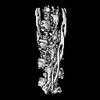
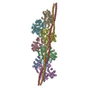
| Title | Structure of the native myosin filament in the relaxed cardiac sarcomere. |
|---|---|
| Journal, issue, pages | Nature, Vol. 623, Issue 7988, Page 863-871, Year 2023 |
| Publish date | Nov 1, 2023 |
 Authors Authors | Davide Tamborrini / Zhexin Wang / Thorsten Wagner / Sebastian Tacke / Markus Stabrin / Michael Grange / Ay Lin Kho / Martin Rees / Pauline Bennett / Mathias Gautel / Stefan Raunser /   |
| PubMed Abstract | The thick filament is a key component of sarcomeres, the basic units of striated muscle. Alterations in thick filament proteins are associated with familial hypertrophic cardiomyopathy and other ...The thick filament is a key component of sarcomeres, the basic units of striated muscle. Alterations in thick filament proteins are associated with familial hypertrophic cardiomyopathy and other heart and muscle diseases. Despite the central importance of the thick filament, its molecular organization remains unclear. Here we present the molecular architecture of native cardiac sarcomeres in the relaxed state, determined by cryo-electron tomography. Our reconstruction of the thick filament reveals the three-dimensional organization of myosin, titin and myosin-binding protein C (MyBP-C). The arrangement of myosin molecules is dependent on their position along the filament, suggesting specialized capacities in terms of strain susceptibility and force generation. Three pairs of titin-α and titin-β chains run axially along the filament, intertwining with myosin tails and probably orchestrating the length-dependent activation of the sarcomere. Notably, whereas the three titin-α chains run along the entire length of the thick filament, titin-β chains do not. The structure also demonstrates that MyBP-C bridges thin and thick filaments, with its carboxy-terminal region binding to the myosin tails and directly stabilizing the OFF state of the myosin heads in an unforeseen manner. These results provide a foundation for future research investigating muscle disorders involving sarcomeric components. |
 External links External links | Nature / PubMed:37914933 / PubMed Central |
| Methods | EM (subtomogram averaging) / EM (tomography) |
| Resolution | 8.0 - 23.6 Å |
| Structure data |  EMDB-16986: Structure of the relaxed thin filament from FIB milled left ventricular mouse myofibrils (tropomyosin masked out)  EMDB-16987: Structure of the relaxed thin filament from FIB milled left ventricular mouse myofibrils (including tropomyosin)  EMDB-16988: Tomogram of sarcomere C-zone from mouse cardiac muscle  EMDB-16989: Tomogram of sarcomere M-band to C-zone from mouse cardiac muscle  EMDB-16990: Structure of the relaxed thick filament from FIB milled left ventricular mouse myofibrils - Crowns P2-A1  EMDB-16991: Structure of the relaxed thick filament from FIB milled left ventricular mouse myofibrils - M-band  EMDB-16992: Structure of the relaxed thick filament from FIB milled left ventricular mouse myofibrils - Crowns A15-A29  EMDB-16993: Structure of the relaxed thick filament from FIB milled left ventricular mouse myofibrils - Crown P1  EMDB-16994: Structure of the relaxed thick filament from FIB milled left ventricular mouse myofibrils - Crowns A11-A15  EMDB-16995: Structure of the relaxed thick filament from FIB milled left ventricular mouse myofibrils - Crowns A8-A12  EMDB-16996: Structure of the relaxed thick filament from FIB milled left ventricular mouse myofibrils - Crowns A5-A7  EMDB-16997: Structure of the relaxed thick filament from FIB milled left ventricular mouse myofibrils - Crowns A1-A5  EMDB-18146: In situ structures from relaxed cardiac myofibrils reveal the organization of the muscle thick filament EMDB-18147, PDB-8q4g: EMDB-18198, PDB-8q6t:  EMDB-18200: Thin filament consensus map from FIB milled relaxed left ventricular mouse myofibrils |
| Source |
|
 Keywords Keywords | STRUCTURAL PROTEIN / muscle sarcomere calcium-free cardiac thin-filament / MOTOR PROTEIN / Mammalian / Muscle / Thick filament / Cardiac |