 Movie
Movie Controller

[English] 日本語
 Yorodumi
- EMDB-18147: Thin filament from FIB milled relaxed left ventricular mouse myof... -
+ Open data

- Basic information
| Entry | 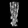 | ||||||||||||
|---|---|---|---|---|---|---|---|---|---|---|---|---|---|
| Title | Thin filament from FIB milled relaxed left ventricular mouse myofibrils | ||||||||||||
 Map data Map data | Composite map (EMD-16986 and EMD-16987) for the thin filament in relaxed ventricular mouse myofibrils. Enhanced with LocSpiral. | ||||||||||||
 Sample Sample |
| ||||||||||||
 Keywords Keywords | muscle sarcomere calcium-free cardiac thin-filament / STRUCTURAL PROTEIN | ||||||||||||
| Function / homology |  Function and homology information Function and homology informationpositive regulation of heart rate by epinephrine / muscle thin filament tropomyosin / RHOB GTPase cycle / Formation of the dystrophin-glycoprotein complex (DGC) / Striated Muscle Contraction / Regulation of CDH1 Function / RHOA GTPase cycle / cytoplasmic actin-based contraction involved in cell motility / Smooth Muscle Contraction / actin-myosin filament sliding ...positive regulation of heart rate by epinephrine / muscle thin filament tropomyosin / RHOB GTPase cycle / Formation of the dystrophin-glycoprotein complex (DGC) / Striated Muscle Contraction / Regulation of CDH1 Function / RHOA GTPase cycle / cytoplasmic actin-based contraction involved in cell motility / Smooth Muscle Contraction / actin-myosin filament sliding / cardiac myofibril assembly / actin filament-based movement / bleb / ruffle organization / cardiac muscle tissue morphogenesis / actomyosin structure organization / actin filament capping / muscle filament sliding / I band / sarcomere organization / ventricular cardiac muscle tissue morphogenesis / microfilament motor activity / myosin binding / heart contraction / negative regulation of vascular associated smooth muscle cell migration / mesenchyme migration / myofibril / negative regulation of vascular associated smooth muscle cell proliferation / skeletal muscle thin filament assembly / cytoskeletal protein binding / positive regulation of stress fiber assembly / cardiac muscle contraction / stress fiber / positive regulation of cell adhesion / negative regulation of cell migration / actin filament organization / sarcomere / filopodium / actin filament / wound healing / structural constituent of cytoskeleton / Hydrolases; Acting on acid anhydrides; Acting on acid anhydrides to facilitate cellular and subcellular movement / ruffle membrane / disordered domain specific binding / actin filament binding / regulation of cell shape / lamellipodium / actin cytoskeleton / actin binding / cell body / in utero embryonic development / response to ethanol / response to xenobiotic stimulus / protein heterodimerization activity / hydrolase activity / positive regulation of gene expression / synapse / negative regulation of apoptotic process / glutamatergic synapse / protein homodimerization activity / protein-containing complex / ATP binding / identical protein binding / cytoplasm / cytosol Similarity search - Function | ||||||||||||
| Biological species |  | ||||||||||||
| Method | subtomogram averaging / cryo EM / Resolution: 8.0 Å | ||||||||||||
 Authors Authors | Tamborrini D / Wang Z / Wagner T / Tacke S / Stabrin M / Grange M / Kho AL / Bennet P / Rees M / Gautel M / Raunser S | ||||||||||||
| Funding support | European Union,  Canada, 3 items Canada, 3 items
| ||||||||||||
 Citation Citation | Journal: Nature / Year: 2023 Title: Structure of the native myosin filament in the relaxed cardiac sarcomere. Authors: Davide Tamborrini / Zhexin Wang / Thorsten Wagner / Sebastian Tacke / Markus Stabrin / Michael Grange / Ay Lin Kho / Martin Rees / Pauline Bennett / Mathias Gautel / Stefan Raunser /   Abstract: The thick filament is a key component of sarcomeres, the basic units of striated muscle. Alterations in thick filament proteins are associated with familial hypertrophic cardiomyopathy and other ...The thick filament is a key component of sarcomeres, the basic units of striated muscle. Alterations in thick filament proteins are associated with familial hypertrophic cardiomyopathy and other heart and muscle diseases. Despite the central importance of the thick filament, its molecular organization remains unclear. Here we present the molecular architecture of native cardiac sarcomeres in the relaxed state, determined by cryo-electron tomography. Our reconstruction of the thick filament reveals the three-dimensional organization of myosin, titin and myosin-binding protein C (MyBP-C). The arrangement of myosin molecules is dependent on their position along the filament, suggesting specialized capacities in terms of strain susceptibility and force generation. Three pairs of titin-α and titin-β chains run axially along the filament, intertwining with myosin tails and probably orchestrating the length-dependent activation of the sarcomere. Notably, whereas the three titin-α chains run along the entire length of the thick filament, titin-β chains do not. The structure also demonstrates that MyBP-C bridges thin and thick filaments, with its carboxy-terminal region binding to the myosin tails and directly stabilizing the OFF state of the myosin heads in an unforeseen manner. These results provide a foundation for future research investigating muscle disorders involving sarcomeric components. | ||||||||||||
| History |
|
- Structure visualization
| Supplemental images |
|---|

- Downloads & links
-EMDB archive
| Map data | emd_18147.map.gz | 492.3 KB | EMDB map data format | |
|---|---|---|---|---|
| Header (meta data) | emd-18147-v30.xmlemd-18147.xml | 13.9 KB 13.9 KB | Display Display | EMDB header |
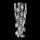
| Images |  emd_18147.png emd_18147.png | 60.9 KB | ||
| Filedesc metadata | emd-18147.cif.gz | 5.8 KB | ||
| Archive directory |  http://ftp.pdbj.org/pub/emdb/structures/EMD-18147ftp://ftp.pdbj.org/pub/emdb/structures/EMD-18147 http://ftp.pdbj.org/pub/emdb/structures/EMD-18147ftp://ftp.pdbj.org/pub/emdb/structures/EMD-18147 | HTTPS FTP |
-Related structure data
| Related structure data |  8q4gMC  8q6tC C: citing same article ( M: atomic model generated by this map |
|---|---|
| Similar structure data |


-Links
| EMDB pages | EMDB (EBI/PDBe) / EMDataResource |
|---|---|
| Related items in Molecule of the Month |


-Map
| File | Download / File: emd_18147.map.gz / Format: CCP4 / Size: 8 MB / Type: IMAGE STORED AS FLOATING POINT NUMBER (4 BYTES) | ||||||||||||||||||||||||||||||||||||
|---|---|---|---|---|---|---|---|---|---|---|---|---|---|---|---|---|---|---|---|---|---|---|---|---|---|---|---|---|---|---|---|---|---|---|---|---|---|
| Annotation | Composite map (EMD-16986 and EMD-16987) for the thin filament in relaxed ventricular mouse myofibrils. Enhanced with LocSpiral. | ||||||||||||||||||||||||||||||||||||
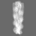
| Projections & slices | Image control
Images are generated by Spider. | ||||||||||||||||||||||||||||||||||||
| Voxel size | X=Y=Z: 2.293 Å | ||||||||||||||||||||||||||||||||||||
| Density |
| ||||||||||||||||||||||||||||||||||||
| Symmetry | Space group: 1 | ||||||||||||||||||||||||||||||||||||
| Details | EMDB XML:
|
 Z (Sec.)
 Y (Row.)
 X (Col.)


-Supplemental data
- Sample components
-Entire : Calcium-free thin filament from relaxed mouse left-ventricular my...
| Entire | Name: Calcium-free thin filament from relaxed mouse left-ventricular myofibrils |
|---|---|
| Components |
|
-Supramolecule #1: Calcium-free thin filament from relaxed mouse left-ventricular my...
| Supramolecule | Name: Calcium-free thin filament from relaxed mouse left-ventricular myofibrils type: tissue / ID: 1 / Parent: 0 / Macromolecule list: #2, #1 |
|---|---|
| Source (natural) | Organism: |
-Macromolecule #1: Actin, alpha cardiac muscle 1
| Macromolecule | Name: Actin, alpha cardiac muscle 1 / type: protein_or_peptide / ID: 1 / Number of copies: 7 / Enantiomer: LEVO EC number: Hydrolases; Acting on acid anhydrides; Acting on acid anhydrides to facilitate cellular and subcellular movement |
|---|---|
| Source (natural) | Organism: |
| Molecular weight | Theoretical: 41.471258 KDa |
| Sequence | String: ETTALVCDNG SGLVKAGFAG DDAPRAVFPS IVGRPRHQGV MVGMGQKDSY VGDEAQSKRG ILTLKYPIEH GIITNWDDME KIWHHTFYN ELRVAPEEHP TLLTEAPLNP KANREKMTQI MFETFNVPAM YVAIQAVLSL YASGRTTGIV LDSGDGVTHN V PIYEGYAL ...String: ETTALVCDNG SGLVKAGFAG DDAPRAVFPS IVGRPRHQGV MVGMGQKDSY VGDEAQSKRG ILTLKYPIEH GIITNWDDME KIWHHTFYN ELRVAPEEHP TLLTEAPLNP KANREKMTQI MFETFNVPAM YVAIQAVLSL YASGRTTGIV LDSGDGVTHN V PIYEGYAL PHAIMRLDLA GRDLTDYLMK ILTERGYSFV TTAEREIVRD IKEKLCYVAL DFENEMATAA SSSSLEKSYE LP DGQVITI GNERFRCPET LFQPSFIGME SAGIHETTYN SIMKCDIDIR KDLYANNVLS GGTTMYPGIA DRMQKEITAL APS TMKIKI IAPPERKYSV WIGGSILASL STFQQMWISK QEYDEAGPSI VHRKCF UniProtKB: Actin, alpha cardiac muscle 1 |
-Macromolecule #2: Tropomyosin alpha-1 chain
| Macromolecule | Name: Tropomyosin alpha-1 chain / type: protein_or_peptide / ID: 2 / Number of copies: 2 / Enantiomer: LEVO |
|---|---|
| Source (natural) | Organism: |
| Molecular weight | Theoretical: 20.840275 KDa |
| Sequence | String: IQLVEEELDR AQERLATALQ KLEEAEKAAD ESERGMKVIE SRAQKDEEKM EIQEIQLKEA KHIAEDADRK YEEVARKLVI IESDLERAE ERAELSEGKC AELEEELKTV TNNLKSLEAQ AEKYSQKEDK YEEEIKVLSD KLKEAETRAE FAERSVTKLE K SIDDLEDE LYAQKLKYKA I UniProtKB: Tropomyosin alpha-1 chain |
-Experimental details
-Structure determination
| Method | cryo EM |
|---|---|
 Processing Processing | subtomogram averaging |
| Aggregation state | tissue |
-Sample preparation
| Buffer | pH: 7 |
|---|---|
| Vitrification | Cryogen name: ETHANE-PROPANE |
- Electron microscopy
| Microscope | FEI TITAN KRIOS |
|---|---|
| Image recording | Film or detector model: GATAN K3 BIOQUANTUM (6k x 4k) / Average electron dose: 3.4 e/Å2 |
| Electron beam | Acceleration voltage: 300 kV / Electron source:  FIELD EMISSION GUN FIELD EMISSION GUN |
| Electron optics | Illumination mode: FLOOD BEAM / Imaging mode: BRIGHT FIELD / Nominal defocus max: 6.0 µm / Nominal defocus min: 3.0 µm / Nominal magnification: 81000 |
| Sample stage | Specimen holder model: FEI TITAN KRIOS AUTOGRID HOLDER |
| Experimental equipment |  Model: Titan Krios / Image courtesy: FEI Company |
-Image processing
| Final reconstruction | Applied symmetry - Point group: C1 (asymmetric) / Resolution.type: BY AUTHOR / Resolution: 8.0 Å / Resolution method: FSC 0.143 CUT-OFF / Software - Name: RELION / Number subtomograms used: 100447 |
|---|---|
| Extraction | Number tomograms: 89 / Number images used: 365971 / Software - Name: Warp |
| Final angle assignment | Type: MAXIMUM LIKELIHOOD / Software - Name: RELION |
-Atomic model buiding 1
| Initial model | Chain - Source name: AlphaFold / Chain - Initial model type: in silico model |
|---|---|
| Output model | PDB-8q4g: |
