Journal: J Struct Biol / Year: 2021 Title: Comparative structural, biophysical, and receptor binding study of true type and wild type AAV2. Authors: Antonette Bennett / Joshua Hull / Nelly Jolinon / Julie Tordo / Katie Moss / Enswert Binns / Mario Mietzsch / Cathleen Hagemann / R Michael Linden / Andrea Serio / Paul Chipman / Duncan ...Authors: Antonette Bennett / Joshua Hull / Nelly Jolinon / Julie Tordo / Katie Moss / Enswert Binns / Mario Mietzsch / Cathleen Hagemann / R Michael Linden / Andrea Serio / Paul Chipman / Duncan Sousa / Felix Broecker / Peter Seeberger / Els Henckaerts / Robert McKenna / Mavis Agbandje-McKenna / Abstract: Adeno-associated viruses (AAV) are utilized as gene transfer vectors in the treatment of monogenic disorders. A variant, rationally engineered based on natural AAV2 isolates, designated AAV-True Type ...Adeno-associated viruses (AAV) are utilized as gene transfer vectors in the treatment of monogenic disorders. A variant, rationally engineered based on natural AAV2 isolates, designated AAV-True Type (AAV-TT), is highly neurotropic compared to wild type AAV2 in vivo, and vectors based on it, are currently being evaluated for central nervous system applications. AAV-TT differs from AAV2 by 14 amino acids, including R585S and R588T, two residues previously shown to be essential for heparan sulfate binding of AAV2. The capsid structures of AAV-TT and AAV2 visualized by cryo-electron microscopy at 3.4 and 3.0 Å resolution, respectively, highlighted structural perturbations at specific amino acid differences. Differential scanning fluorimetry (DSF) performed at different pH conditions demonstrated that the melting temperature (T) of AAV2 was consistently ∼5 °C lower than AAV-TT, but both showed maximal stability at pH 5.5, corresponding to the pH in the late endosome, proposed as required for VP1u externalization to facilitate endosomal escape. Reintroduction of arginines at positions 585 and 588 in AAV-TT caused a reduction in T, demonstrating that the lack of basic amino acids at these positions are associated with capsid stability. These results provide structural and thermal annotation of AAV2/AAV-TT residue differences, that account for divergent cell binding, transduction, antigenic reactivity, and transduction of permissive tissues between the two viruses. Specifically, these data indicate that AAV-TT may not utilize a glycan receptor mediated pathway to enter cells and may have lower antigenic properties as compared to AAV2.
#200 - Aug 2016 Quasisymmetry in Icosahedral Viruses similarity (1)
-
Assembly
Deposited unit
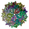
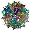
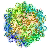
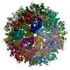
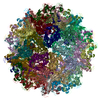
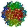
A: Capsid protein VP1 B: Capsid protein VP1 C: Capsid protein VP1 D: Capsid protein VP1 E: Capsid protein VP1 F: Capsid protein VP1 G: Capsid protein VP1 H: Capsid protein VP1 I: Capsid protein VP1 J: Capsid protein VP1 K: Capsid protein VP1 L: Capsid protein VP1 M: Capsid protein VP1 N: Capsid protein VP1 O: Capsid protein VP1 P: Capsid protein VP1 Q: Capsid protein VP1 R: Capsid protein VP1 S: Capsid protein VP1 T: Capsid protein VP1 U: Capsid protein VP1 V: Capsid protein VP1 W: Capsid protein VP1 X: Capsid protein VP1 Y: Capsid protein VP1 Z: Capsid protein VP1 a: Capsid protein VP1 b: Capsid protein VP1 c: Capsid protein VP1 d: Capsid protein VP1 e: Capsid protein VP1 f: Capsid protein VP1 g: Capsid protein VP1 h: Capsid protein VP1 i: Capsid protein VP1 j: Capsid protein VP1 k: Capsid protein VP1 l: Capsid protein VP1 m: Capsid protein VP1 n: Capsid protein VP1 o: Capsid protein VP1 p: Capsid protein VP1 q: Capsid protein VP1 r: Capsid protein VP1 s: Capsid protein VP1 t: Capsid protein VP1 u: Capsid protein VP1 v: Capsid protein VP1 w: Capsid protein VP1 x: Capsid protein VP1 y: Capsid protein VP1 z: Capsid protein VP1 1: Capsid protein VP1 2: Capsid protein VP1 3: Capsid protein VP1 4: Capsid protein VP1 5: Capsid protein VP1 6: Capsid protein VP1 7: Capsid protein VP1 8: Capsid protein VP1
In the structure databanks used in Yorodumi, some data are registered as the other names, "COVID-19 virus" and "2019-nCoV". Here are the details of the virus and the list of structure data.
Jan 31, 2019. EMDB accession codes are about to change! (news from PDBe EMDB page)
EMDB accession codes are about to change! (news from PDBe EMDB page)
The allocation of 4 digits for EMDB accession codes will soon come to an end. Whilst these codes will remain in use, new EMDB accession codes will include an additional digit and will expand incrementally as the available range of codes is exhausted. The current 4-digit format prefixed with “EMD-” (i.e. EMD-XXXX) will advance to a 5-digit format (i.e. EMD-XXXXX), and so on. It is currently estimated that the 4-digit codes will be depleted around Spring 2019, at which point the 5-digit format will come into force.
The EM Navigator/Yorodumi systems omit the EMD- prefix.
Related info.:Q: What is EMD? / ID/Accession-code notation in Yorodumi/EM Navigator
Yorodumi is a browser for structure data from EMDB, PDB, SASBDB, etc.
This page is also the successor to EM Navigator detail page, and also detail information page/front-end page for Omokage search.
The word "yorodu" (or yorozu) is an old Japanese word meaning "ten thousand". "mi" (miru) is to see.
Related info.:EMDB / PDB / SASBDB / Comparison of 3 databanks / Yorodumi Search / Aug 31, 2016. New EM Navigator & Yorodumi / Yorodumi Papers / Jmol/JSmol / Function and homology information / Changes in new EM Navigator and Yorodumi
 Movie
Movie Controller


 Open data
 Basic information
 Components
 Keywords
 Function and homology information
 Adeno-associated virus
 Authors
 Citation


 Structure visualization
 Downloads & links
 Other downloads
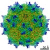


 PDBj
PDBj


 Assembly

 Spodoptera frugiperda (fall armyworm) / References: UniProt: Q670S0
 Sample preparation
 Electron microscopy imaging
 FIELD EMISSION GUN / Accelerating voltage: 300 kV / Illumination mode: FLOOD BEAM
 Processing