Movie
Movie Controller


+ Open data

- Basic information
| Entry | Database: PDB / ID: 7mt0 | |||||||||||||||||||||||||||||||||
|---|---|---|---|---|---|---|---|---|---|---|---|---|---|---|---|---|---|---|---|---|---|---|---|---|---|---|---|---|---|---|---|---|---|---|
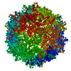
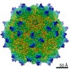
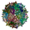
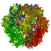
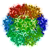
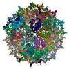
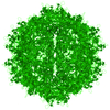
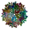
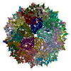
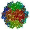
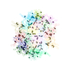
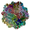
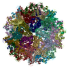
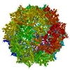
| Title | Structure of the adeno-associated virus 9 capsid at pH 7.4 | |||||||||||||||||||||||||||||||||
 Components Components | Capsid protein VP1 | |||||||||||||||||||||||||||||||||
 Keywords Keywords | VIRUS LIKE PARTICLE / adeno-associated virus / pH / phospholipase-A2 / capsid / gene therapy / AAV9 / dependoparvovirus / intracellular trafficking / endosome | |||||||||||||||||||||||||||||||||
| Function / homology | Phospholipase A2-like domain / Phospholipase A2-like domain / Parvovirus coat protein VP2 / Parvovirus coat protein VP1/VP2 / Parvovirus coat protein VP1/VP2 / Capsid/spike protein, ssDNA virus / T=1 icosahedral viral capsid / structural molecule activity / Capsid protein VP1 Function and homology information Function and homology information | |||||||||||||||||||||||||||||||||
| Biological species |   Adeno-associated virus 9 Adeno-associated virus 9 | |||||||||||||||||||||||||||||||||
| Method | ELECTRON MICROSCOPY / single particle reconstruction / cryo EM / Resolution: 2.82 Å | |||||||||||||||||||||||||||||||||
 Authors Authors | Penzes, J.J. / Chipman, P. / Bhattacharya, N. / Zeher, A. / Huang, R. / McKenna, R. / Agbandje-McKenna, M. | |||||||||||||||||||||||||||||||||
| Funding support |  United States, 5items United States, 5items
| |||||||||||||||||||||||||||||||||
 Citation Citation | Journal: J Virol / Year: 2021 Title: Adeno-associated Virus 9 Structural Rearrangements Induced by Endosomal Trafficking pH and Glycan Attachment. Authors: Judit J Penzes / Paul Chipman / Nilakshee Bhattacharya / Allison Zeher / Rick Huang / Robert McKenna / Mavis Agbandje-McKenna / Abstract: Adeno-associated viruses (AAVs) are small nonenveloped single-stranded DNA (ssDNA) viruses that are currently being developed as gene therapy biologics. After cell entry, AAVs traffic to the nucleus ...Adeno-associated viruses (AAVs) are small nonenveloped single-stranded DNA (ssDNA) viruses that are currently being developed as gene therapy biologics. After cell entry, AAVs traffic to the nucleus using the endo-lysosomal pathway. The subsequent decrease in pH triggers conformational changes to the capsid that enable the externalization of the capsid protein (VP) N termini, including the unique domain of the minor capsid protein VP1 (VP1u), which permits the phospholipase activity required for the capsid lysosomal egress. Here, we report the AAV9 capsid structure, determined at the endosomal pHs (7.4, 6.0, 5.5, and 4.0), and terminal galactose-bound AAV9 capsids at pHs 7.4 and 5.5 using cryo-electron microscopy and three-dimensional image reconstruction. Taken together, these studies provide insight into AAV9 capsid conformational changes at the 5-fold pore during endosomal trafficking, in both the presence and absence of its cellular glycan receptor. We visualized, for the first time, that acidification induces the externalization of the VP3 and possibly VP2 N termini, presumably in prelude to the externalization of VP1u at pH 4.0, which is essential for lysosomal membrane disruption. In addition, the structural study of AAV9-galactose interactions demonstrates that AAV9 remains attached to its glycan receptor at the late endosome pH 5.5. This interaction significantly alters the conformational stability of the variable region I of the VPs, as well as the dynamics associated with VP N terminus externalization. There are 13 distinct Adeno-associated virus (AAV) serotypes that are structurally homologous and whose capsid proteins (VP1 to -3) are similar in amino acid sequence. However, AAV9 is one of the most commonly studied and is used as a gene therapy vector. This is partly because AAV9 is capable of crossing the blood-brain barrier and readily transduces a wide array of tissues, including the central nervous system. In this study, we provide AAV9 capsid structural insight during intracellular trafficking. Although the AAV capsid has been shown to externalize the N termini of its VPs, to enzymatically disrupt the lysosome membrane at low pH, there was no structural evidence to confirm this. By utilizing AAV9 as our model, we provide the first structural evidence that the externalization process occurs at the protein interface at the icosahedral 5-fold symmetry axis and can be triggered by lowering the pH. | |||||||||||||||||||||||||||||||||
| History |
|
- Structure visualization
| Movie |
Movie viewer |
|---|---|
| Structure viewer | Molecule: MolmilJmol/JSmol |
 UCSF Chimera
- Downloads & links
-Download
| PDBx/mmCIF format | 7mt0.cif.gz | 5.6 MB | Display | PDBx/mmCIF format |
|---|---|---|---|---|
| PDB format | pdb7mt0.ent.gz | Display | PDB format | |
| PDBx/mmJSON format | 7mt0.json.gz | Tree view | PDBx/mmJSON format | |
| Others |  Other downloads Other downloads |
-Validation report
| Arichive directory | https://data.pdbj.org/pub/pdb/validation_reports/mt/7mt0ftp://data.pdbj.org/pub/pdb/validation_reports/mt/7mt0 | HTTPS FTP |
|---|
-Related structure data
| Related structure data | 23973MC  7mtgC  7mtpC  7mtwC  7mtzC  7muaC M: map data used to model this data C: citing same article ( |
|---|---|
| Similar structure data |


-Links
 PDBj
PDBj


- Assembly
| Deposited unit | 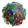
|
|---|---|
| 1 |
|
-Components
| #1: Protein | Mass: 58474.414 Da / Num. of mol.: 60 / Fragment: UNP residues 219-736 Source method: isolated from a genetically manipulated source Source: (gene. exp.) Adeno-associated virus 9 / Gene: cap / Plasmid: pFastBac1-HM / Cell line (production host): Sf9 / Production host:   Spodoptera frugiperda (fall armyworm) / References: UniProt: Q6JC40 Spodoptera frugiperda (fall armyworm) / References: UniProt: Q6JC40Has protein modification | N | |
|---|
-Experimental details
-Experiment
| Experiment | Method: ELECTRON MICROSCOPY |
|---|---|
| EM experiment | Aggregation state: PARTICLE / 3D reconstruction method: single particle reconstruction |
- Sample preparation
| Component | Name: Adeno-associated virus 9 / Type: VIRUS / Entity ID: all / Source: RECOMBINANT |
|---|---|
| Molecular weight | Experimental value: NO |
| Source (natural) | Organism: Adeno-associated virus 9 |
| Source (recombinant) | Organism: Spodoptera frugiperda (fall armyworm) / Cell: Sf9 / Plasmid: pFastBac1-HM |
| Details of virus | Empty: YES / Enveloped: NO / Isolate: SEROTYPE / Type: VIRUS-LIKE PARTICLE |
| Natural host | Organism: Homo sapiens |
| Virus shell | Name: AAV9 virus like particle / Diameter: 240 nm / Triangulation number (T number): 1 |
| Buffer solution | pH: 7.4 Details: 1x Universal buffer of 20 mM Hepes, 20 mM MES, 20 mM sodium acetate, 0.15 M NaCl, 3.7 mM CaCl2 |
| Specimen | Embedding applied: NO / Shadowing applied: NO / Staining applied: NO / Vitrification applied: YES |
| Specimen support | Grid material: COPPER / Grid type: Quantifoil |
| Vitrification | Cryogen name: ETHANE |
- Electron microscopy imaging
| Experimental equipment |  Model: Titan Krios / Image courtesy: FEI Company |
|---|---|
| Microscopy | Model: FEI TITAN KRIOS |
| Electron gun | Electron source: OTHER / Accelerating voltage: 300 kV / Illumination mode: FLOOD BEAM |
| Electron lens | Mode: OTHER / Cs: 2.7 mm |
| Image recording | Electron dose: 60 e/Å2 / Film or detector model: GATAN K2 SUMMIT (4k x 4k) |
- Processing
| Software | Name: PHENIX / Version: 1.10-2155_2155: / Classification: refinement | |||||||||||||||||||||||||||||||||||
|---|---|---|---|---|---|---|---|---|---|---|---|---|---|---|---|---|---|---|---|---|---|---|---|---|---|---|---|---|---|---|---|---|---|---|---|---|
| EM software |
| |||||||||||||||||||||||||||||||||||
| CTF correction | Type: PHASE FLIPPING ONLY | |||||||||||||||||||||||||||||||||||
| Symmetry | Point symmetry: I (icosahedral) | |||||||||||||||||||||||||||||||||||
| 3D reconstruction | Resolution: 2.82 Å / Resolution method: FSC 0.143 CUT-OFF / Num. of particles: 150469 / Algorithm: FOURIER SPACE / Symmetry type: POINT | |||||||||||||||||||||||||||||||||||
| Atomic model building | Protocol: RIGID BODY FIT / Space: REAL | |||||||||||||||||||||||||||||||||||
| Atomic model building | PDB-ID: 3UX1 Pdb chain-ID: A / Accession code: 3UX1 / Source name: PDB / Type: experimental model | |||||||||||||||||||||||||||||||||||
| Refine LS restraints |
|