 Movie
Movie Controller

[English] 日本語
 Yorodumi
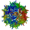
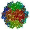
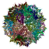
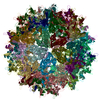
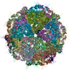
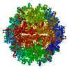
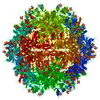
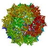
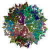
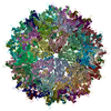
- PDB-6cbe: Atomic structure of a rationally engineered gene delivery vector,... -
+ Open data

- Basic information
| Entry | Database: PDB / ID: 6cbe | |||||||||||||||||||||||||||||||||
|---|---|---|---|---|---|---|---|---|---|---|---|---|---|---|---|---|---|---|---|---|---|---|---|---|---|---|---|---|---|---|---|---|---|---|
| Title | Atomic structure of a rationally engineered gene delivery vector, AAV2.5 | |||||||||||||||||||||||||||||||||
 Components Components | Capsid protein VP1 | |||||||||||||||||||||||||||||||||
 Keywords Keywords | VIRUS / AAV / dependoparvovirus / gene therapy vector / retional design | |||||||||||||||||||||||||||||||||
| Function / homology |  Function and homology information Function and homology informationsymbiont entry into host cell via permeabilization of host membrane / host cell nucleolus / T=1 icosahedral viral capsid / clathrin-dependent endocytosis of virus by host cell / virion attachment to host cell / structural molecule activity Similarity search - Function | |||||||||||||||||||||||||||||||||
| Biological species |  Adeno-associated virus 2 Adeno-associated virus 2 | |||||||||||||||||||||||||||||||||
| Method | ELECTRON MICROSCOPY / single particle reconstruction / cryo EM / Resolution: 2.78 Å | |||||||||||||||||||||||||||||||||
 Authors Authors | Burg, M. / Rosebrough, C. / Drouin, L. / Bennett, A. / Mietzsch, M. / Chipman, P. / McKenna, R. / Sousa, D. / Potter, M. / Byrne, B. ...Burg, M. / Rosebrough, C. / Drouin, L. / Bennett, A. / Mietzsch, M. / Chipman, P. / McKenna, R. / Sousa, D. / Potter, M. / Byrne, B. / Kozyreva, O.G. / Samulski, R.J. / Agbandje-McKenna, M. | |||||||||||||||||||||||||||||||||
 Citation Citation | Journal: J Struct Biol / Year: 2018 Title: Atomic structure of a rationally engineered gene delivery vector, AAV2.5. Authors: Matthew Burg / Claire Rosebrough / Lauren M Drouin / Antonette Bennett / Mario Mietzsch / Paul Chipman / Robert McKenna / Duncan Sousa / Mark Potter / Barry Byrne / R Jude Samulski / Mavis Agbandje-McKenna /  Abstract: AAV2.5 represents the first structure-guided in-silico designed Adeno-associated virus (AAV) gene delivery vector. This engineered vector combined the receptor attachment properties of AAV serotype 2 ...AAV2.5 represents the first structure-guided in-silico designed Adeno-associated virus (AAV) gene delivery vector. This engineered vector combined the receptor attachment properties of AAV serotype 2 (AAV2) with the muscle tropic properties of AAV1, and exhibited an antibody escape phenotype because of a modified antigenic epitope. To confirm the design, the structure of the vector was determined to a resolution of 2.78 Å using cryo-electron microscopy and image reconstruction. The structure of the major viral protein (VP), VP3, was ordered from residue 219 to 736, as reported for other AAV structures, and the five AAV2.5 residues exchanged from AAV2 to AAV1, Q263A, T265 (insertion), N706A, V709A, and T717N, were readily interpretable. Significantly, the surface loops containing these residues adopt the AAV1 conformation indicating the importance of amino acid residues in dictating VP structure. | |||||||||||||||||||||||||||||||||
| History |
|
- Structure visualization
| Movie |
Movie viewer |
|---|---|
| Structure viewer | Molecule: MolmilJmol/JSmol |
- Downloads & links
-Download
| PDBx/mmCIF format | 6cbe.cif.gz | 5.8 MB | Display | PDBx/mmCIF format |
|---|---|---|---|---|
| PDB format | pdb6cbe.ent.gz | Display | PDB format | |
| PDBx/mmJSON format | 6cbe.json.gz | Tree view | PDBx/mmJSON format | |
| Others |  Other downloads Other downloads |
-Validation report
| Arichive directory | https://data.pdbj.org/pub/pdb/validation_reports/cb/6cbeftp://data.pdbj.org/pub/pdb/validation_reports/cb/6cbe | HTTPS FTP |
|---|
-Related structure data
| Related structure data | 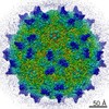 7452MC M: map data used to model this data C: citing same article ( |
|---|---|
| Similar structure data |


-Links
 PDBj
PDBj


- Assembly
| Deposited unit | 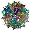
|
|---|---|
| 1 |
|
-Components
| #1: Protein | Mass: 82017.328 Da / Num. of mol.: 60 Source method: isolated from a genetically manipulated source Source: (gene. exp.) Adeno-associated virus 2 / Gene: VP1 / Cell line (production host): HEK293 / Production host:  Homo sapiens (human) / References: UniProt: P03135 Homo sapiens (human) / References: UniProt: P03135Has protein modification | N | |
|---|
-Experimental details
-Experiment
| Experiment | Method: ELECTRON MICROSCOPY |
|---|---|
| EM experiment | Aggregation state: PARTICLE / 3D reconstruction method: single particle reconstruction |
- Sample preparation
| Component | Name: AAV2.5 / Type: VIRUS / Entity ID: all / Source: RECOMBINANT |
|---|---|
| Source (natural) | Organism: Adeno-associated virus 2 |
| Source (recombinant) | Organism: Homo sapiens (human) / Cell: HEK293 |
| Details of virus | Empty: YES / Enveloped: NO / Isolate: OTHER / Type: VIRION |
| Buffer solution | pH: 7.4 |
| Specimen | Embedding applied: NO / Shadowing applied: NO / Staining applied: NO / Vitrification applied: YES |
| Vitrification | Cryogen name: ETHANE |
- Electron microscopy imaging
| Experimental equipment |  Model: Titan Krios / Image courtesy: FEI Company |
|---|---|
| Microscopy | Model: FEI TITAN KRIOS |
| Electron gun | Electron source:  FIELD EMISSION GUN / Accelerating voltage: 300 kV / Illumination mode: FLOOD BEAM FIELD EMISSION GUN / Accelerating voltage: 300 kV / Illumination mode: FLOOD BEAM |
| Electron lens | Mode: BRIGHT FIELD / Cs: 2.7 mm |
| Image recording | Electron dose: 2 e/Å2 / Film or detector model: DIRECT ELECTRON DE-20 (5k x 3k) |
- Processing
| Software | Name: PHENIX / Version: 1.10-2155_2155: / Classification: refinement | ||||||||||||||||||||||||
|---|---|---|---|---|---|---|---|---|---|---|---|---|---|---|---|---|---|---|---|---|---|---|---|---|---|
| EM software |
| ||||||||||||||||||||||||
| CTF correction | Type: PHASE FLIPPING AND AMPLITUDE CORRECTION | ||||||||||||||||||||||||
| 3D reconstruction | Resolution: 2.78 Å / Resolution method: FSC 0.143 CUT-OFF / Num. of particles: 24618 / Symmetry type: POINT | ||||||||||||||||||||||||
| Refine LS restraints |
|
