[English] 日本語
 Yorodumi
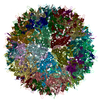
- EMDB-7452: Atomic structure of a rationally engineered gene delivery vector,... -
+ Open data
- Basic information
| Entry | Database: EMDB / ID: EMD-7452 | |||||||||
|---|---|---|---|---|---|---|---|---|---|---|
| Title | Atomic structure of a rationally engineered gene delivery vector, AAV2.5 | |||||||||
 Map data Map data | rationally engineered gene delivery vector, AAV2.5 | |||||||||
 Sample Sample | AAV2.5 != Adeno-associated virus 2 AAV2.5
| |||||||||
 Keywords Keywords | AAV / dependoparvovirus / gene therapy vector / retional design / VIRUS | |||||||||
| Function / homology |  Function and homology information Function and homology informationsymbiont entry into host cell via permeabilization of host membrane / host cell nucleolus / T=1 icosahedral viral capsid / clathrin-dependent endocytosis of virus by host cell / virion attachment to host cell / structural molecule activity Similarity search - Function | |||||||||
| Biological species |  Adeno-associated virus 2 Adeno-associated virus 2 | |||||||||
| Method | single particle reconstruction / cryo EM / Resolution: 2.78 Å | |||||||||
 Authors Authors | Burg M / Rosebrough C | |||||||||
 Citation Citation |  Journal: J Struct Biol / Year: 2018 Journal: J Struct Biol / Year: 2018Title: Atomic structure of a rationally engineered gene delivery vector, AAV2.5. Authors: Matthew Burg / Claire Rosebrough / Lauren M Drouin / Antonette Bennett / Mario Mietzsch / Paul Chipman / Robert McKenna / Duncan Sousa / Mark Potter / Barry Byrne / R Jude Samulski / Mavis Agbandje-McKenna /  Abstract: AAV2.5 represents the first structure-guided in-silico designed Adeno-associated virus (AAV) gene delivery vector. This engineered vector combined the receptor attachment properties of AAV serotype 2 ...AAV2.5 represents the first structure-guided in-silico designed Adeno-associated virus (AAV) gene delivery vector. This engineered vector combined the receptor attachment properties of AAV serotype 2 (AAV2) with the muscle tropic properties of AAV1, and exhibited an antibody escape phenotype because of a modified antigenic epitope. To confirm the design, the structure of the vector was determined to a resolution of 2.78 Å using cryo-electron microscopy and image reconstruction. The structure of the major viral protein (VP), VP3, was ordered from residue 219 to 736, as reported for other AAV structures, and the five AAV2.5 residues exchanged from AAV2 to AAV1, Q263A, T265 (insertion), N706A, V709A, and T717N, were readily interpretable. Significantly, the surface loops containing these residues adopt the AAV1 conformation indicating the importance of amino acid residues in dictating VP structure. | |||||||||
| History |
|
- Structure visualization
| Movie |
 Movie viewer Movie viewer |
|---|---|
| Structure viewer | EM map:  SurfView SurfView Molmil Molmil Jmol/JSmol Jmol/JSmol |
| Supplemental images |
- Downloads & links
-EMDB archive
| Map data |  emd_7452.map.gz emd_7452.map.gz | 84.3 MB |  EMDB map data format EMDB map data format | |
|---|---|---|---|---|
| Header (meta data) |  emd-7452-v30.xml emd-7452-v30.xml emd-7452.xml emd-7452.xml | 12.9 KB 12.9 KB | Display Display |  EMDB header EMDB header |
| Images |  emd_7452.png emd_7452.png | 257.5 KB | ||
| Filedesc metadata |  emd-7452.cif.gz emd-7452.cif.gz | 5.8 KB | ||
| Archive directory |  http://ftp.pdbj.org/pub/emdb/structures/EMD-7452 http://ftp.pdbj.org/pub/emdb/structures/EMD-7452 ftp://ftp.pdbj.org/pub/emdb/structures/EMD-7452 ftp://ftp.pdbj.org/pub/emdb/structures/EMD-7452 | HTTPS FTP |
-Related structure data
| Related structure data |  6cbeMC M: atomic model generated by this map C: citing same article ( |
|---|---|
| Similar structure data |
- Links
Links
| EMDB pages |  EMDB (EBI/PDBe) / EMDB (EBI/PDBe) /  EMDataResource EMDataResource |
|---|---|
| Related items in Molecule of the Month |
- Map
Map
| File |  Download / File: emd_7452.map.gz / Format: CCP4 / Size: 242.3 MB / Type: IMAGE STORED AS FLOATING POINT NUMBER (4 BYTES) Download / File: emd_7452.map.gz / Format: CCP4 / Size: 242.3 MB / Type: IMAGE STORED AS FLOATING POINT NUMBER (4 BYTES) | ||||||||||||||||||||||||||||||||||||||||||||||||||||||||||||||||||||
|---|---|---|---|---|---|---|---|---|---|---|---|---|---|---|---|---|---|---|---|---|---|---|---|---|---|---|---|---|---|---|---|---|---|---|---|---|---|---|---|---|---|---|---|---|---|---|---|---|---|---|---|---|---|---|---|---|---|---|---|---|---|---|---|---|---|---|---|---|---|
| Annotation | rationally engineered gene delivery vector, AAV2.5 | ||||||||||||||||||||||||||||||||||||||||||||||||||||||||||||||||||||
| Projections & slices | Image control
Images are generated by Spider. | ||||||||||||||||||||||||||||||||||||||||||||||||||||||||||||||||||||
| Voxel size | X=Y=Z: 0.95 Å | ||||||||||||||||||||||||||||||||||||||||||||||||||||||||||||||||||||
| Density |
| ||||||||||||||||||||||||||||||||||||||||||||||||||||||||||||||||||||
| Symmetry | Space group: 1 | ||||||||||||||||||||||||||||||||||||||||||||||||||||||||||||||||||||
| Details | EMDB XML:
CCP4 map header:
| ||||||||||||||||||||||||||||||||||||||||||||||||||||||||||||||||||||
-Supplemental data
- Sample components
-Entire : AAV2.5
| Entire | Name: AAV2.5 |
|---|---|
| Components |
|
-Supramolecule #1: Adeno-associated virus 2
| Supramolecule | Name: Adeno-associated virus 2 / type: virus / ID: 1 / Parent: 0 / Macromolecule list: all / NCBI-ID: 10804 / Sci species name: Adeno-associated virus 2 / Virus type: VIRION / Virus isolate: OTHER / Virus enveloped: No / Virus empty: Yes |
|---|
-Macromolecule #1: Capsid protein VP1
| Macromolecule | Name: Capsid protein VP1 / type: protein_or_peptide / ID: 1 / Number of copies: 60 / Enantiomer: LEVO |
|---|---|
| Source (natural) | Organism:  Adeno-associated virus 2 Adeno-associated virus 2 |
| Molecular weight | Theoretical: 82.017328 KDa |
| Recombinant expression | Organism:  Homo sapiens (human) Homo sapiens (human) |
| Sequence | String: MAADGYLPDW LEDTLSEGIR QWWKLKPGPP PPKPAERHKD DSRGLVLPGY KYLGPFNGLD KGEPVNEADA AALEHDKAYD RQLDSGDNP YLKYNHADAE FQERLKEDTS FGGNLGRAVF QAKKRVLEPL GLVEEPVKTA PGKKRPVEHS PVEPDSSSGT G KAGQQPAR ...String: MAADGYLPDW LEDTLSEGIR QWWKLKPGPP PPKPAERHKD DSRGLVLPGY KYLGPFNGLD KGEPVNEADA AALEHDKAYD RQLDSGDNP YLKYNHADAE FQERLKEDTS FGGNLGRAVF QAKKRVLEPL GLVEEPVKTA PGKKRPVEHS PVEPDSSSGT G KAGQQPAR KRLNFGQTGD ADSVPDPQPL GQPPAAPSGL GTNTMATGSG APMADNNEGA DGVGNSSGNW HCDSTWMGDR VI TTSTRTW ALPTYNNHLY KQISSASTGA SNDNHYFGYS TPWGYFDFNR FHCHFSPRDW QRLINNNWGF RPKRLNFKLF NIQ VKEVTQ NDGTTTIANN LTSTVQVFTD SEYQLPYVLG SAHQGCLPPF PADVFMVPQY GYLTLNNGSQ AVGRSSFYCL EYFP SQMLR TGNNFTFSYT FEDVPFHSSY AHSQSLDRLM NPLIDQYLYY LSRTNTPSGT TTQSRLQFSQ AGASDIRDQS RNWLP GPCY RQQRVSKTSA DNNNSEYSWT GATKYHLNGR DSLVNPGPAM ASHKDDEEKF FPQSGVLIFG KQGSEKTNVD IEKVMI TDE EEIRTTNPVA TEQYGSVSTN LQRGNRQAAT ADVNTQGVLP GMVWQDRDVY LQGPIWAKIP HTDGHFHPSP LMGGFGL KH PPPQILIKNT PVPANPSTTF SAAKFASFIT QYSTGQVSVE IEWELQKENS KRWNPEIQYT SNYAKSANVD FTVDNNGV Y SEPRPIGTRY LTRNL UniProtKB: Capsid protein VP1 |
-Experimental details
-Structure determination
| Method | cryo EM |
|---|---|
 Processing Processing | single particle reconstruction |
| Aggregation state | particle |
- Sample preparation
| Buffer | pH: 7.4 |
|---|---|
| Vitrification | Cryogen name: ETHANE |
- Electron microscopy
| Microscope | FEI TITAN KRIOS |
|---|---|
| Image recording | Film or detector model: DIRECT ELECTRON DE-20 (5k x 3k) / Average electron dose: 2.0 e/Å2 |
| Electron beam | Acceleration voltage: 300 kV / Electron source:  FIELD EMISSION GUN FIELD EMISSION GUN |
| Electron optics | Illumination mode: FLOOD BEAM / Imaging mode: BRIGHT FIELD / Cs: 2.7 mm |
| Experimental equipment |  Model: Titan Krios / Image courtesy: FEI Company |
 Movie
Movie Controller


 Z (Sec.)
 X (Row.)
 Y (Col.)


